Brain recordings reveal the secret to visual memory storage
Researchers have discovered how your brain organizes visual memories by category. This new insight could lead to devices that restore lost memory.

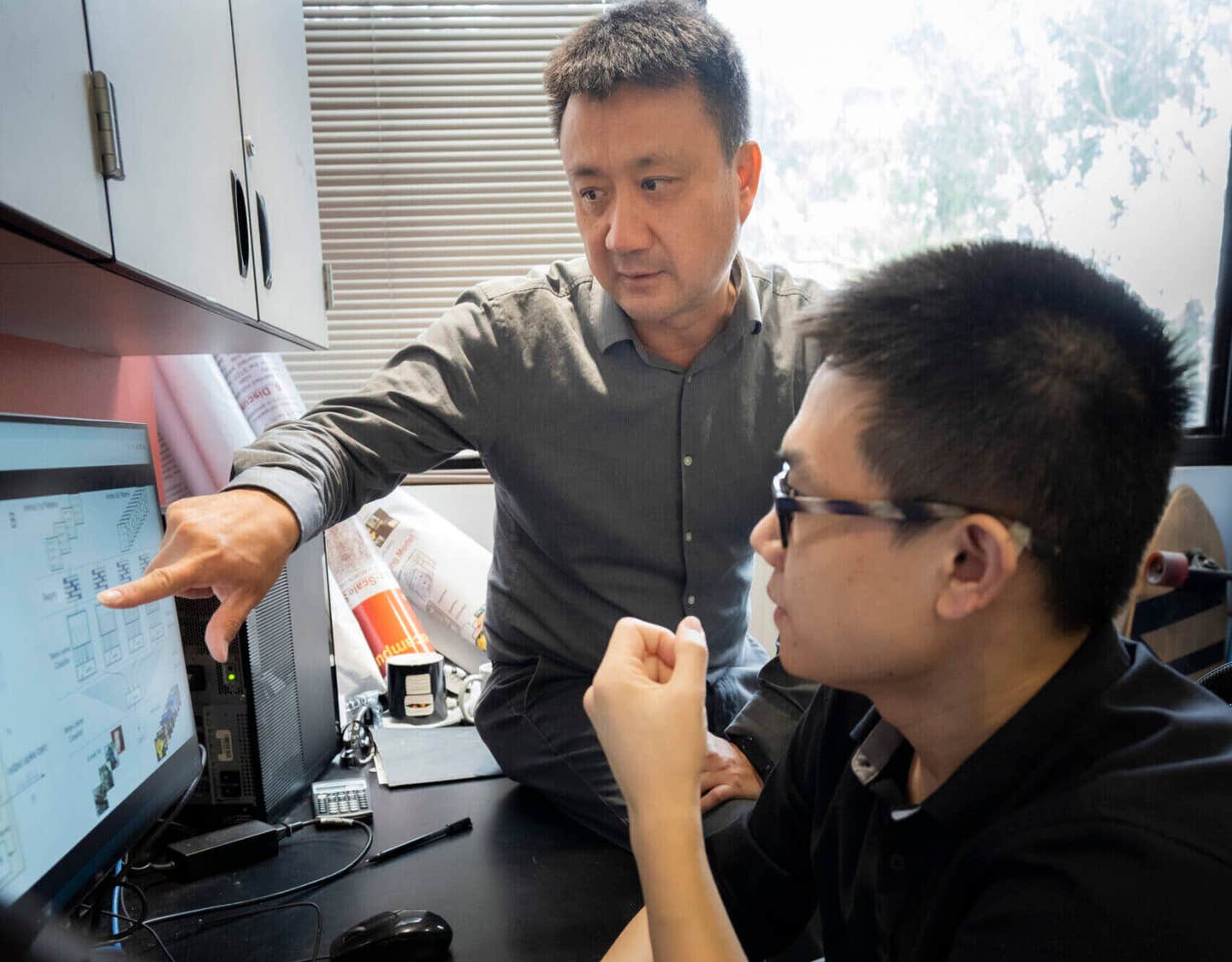
Associate professor Dong Song (L) and first author Xiwei She (R) discuss their machine learning model. (CREDIT: USC)
Scientists have long known that the hippocampus is essential for forming new memories. It helps record where and when things happen. But how it processes what you see—like objects or images—has been a harder mystery to solve. A new study from researchers at the University of Southern California offers a powerful look into this question. Their work shows that the brain may use categories to sort visual memories, making it easier to recall and store them.
Using brain recordings from 24 people with epilepsy, the research team was able to “read” which kind of image a person was remembering based on their brain activity alone. This breakthrough reveals a clear strategy the brain uses to organize complex visual input and may help scientists build memory-restoring devices in the future.
Brain's Filing System for Images
Every day, your brain takes in a flood of images—animals, buildings, tools, plants, and vehicles. Instead of saving every object like a separate photo, the brain may group them into categories. This makes storage more efficient and helps avoid overload.
Researchers have long wondered how the brain manages this task, especially in the hippocampus. This region is well-known for building episodic memories—memories about what happened, where it happened, and when. But how it encodes the "what" of an event, especially with so many objects in the world, has been harder to crack.
Dong Song, an associate professor at USC and director of the Neural Modeling and Interface Laboratory, led the study alongside Charles Liu, director of the USC Neurorestoration Center. The work was published in Advanced Science.
Their team included Xiwei She, the first author and a postdoctoral researcher at Stanford, who developed key parts of the study during his Ph.D. in the Song Lab. Rather than record how a brain responds to a few repeated images, this study looked at how the brain organizes categories of images in real-time. The goal was to see if memory is stored by object type instead of object detail—and how that information is written in the brain’s language.
Decoding the Brain’s Language
To uncover this code, the researchers studied 24 patients with epilepsy. These individuals already had electrodes implanted in their brains to find the source of their seizures. This setup offered a rare chance to listen to neurons in deep areas like the hippocampus.
Related Stories
- Mediterranean diet can slow memory decline and reduce dementia risk
- Breakthrough laser therapy boosts memory recall by 25%
The patients were asked to perform a delayed match-to-sample task. This is a common neuroscience test to study short-term memory. Participants saw an image from one of five categories—animal, plant, building, vehicle, or small tool. After a delay, they had to pick the image they had just seen from a group of options.
While they did this, the team recorded spikes of electrical activity in hippocampal CA1 and CA3 neurons. These spikes are the brain’s way of communicating. By using a machine learning model, the team could analyze the pattern of spikes and decode which image category the patient was remembering. “We let the patients see five categories of images... Then we recorded the hippocampal signal,” Song explained. “Can we decode what category image they are recalling purely based on their brain signal?” The answer turned out to be yes.
How the Hippocampus Encodes Memory
The machine learning model showed that memory categories could be decoded with surprising accuracy. It didn’t rely just on how often a neuron fired, but also when it fired. This type of temporal code—where the timing of spikes carries meaning—is a more advanced way for the brain to store information.
That finding marked a major step forward. Most earlier studies focused only on the average firing rate of neurons. But the USC team discovered that even very brief bursts of activity—on the scale of milliseconds—could signal what kind of image a person was remembering.
This pattern was not limited to a few neurons. In fact, 70% to 80% of the recorded neurons took part in encoding the image categories. But only specific time points within each neuron’s activity were crucial for identifying a category. The brain seems to be using a population code, where many neurons contribute to the message, but each one plays its part in a carefully timed way. “It’s like reading your hippocampus to see what kind of memory you are trying to form,” said Song. “We can pretty accurately decode what kind of category of image the patient was trying to remember.”
Similar Signals in Neighboring Brain Regions
The team also noticed something interesting between the CA1 and CA3 regions of the hippocampus. These two areas showed overlapping, or redundant, memory signals. Both regions could be used to decode the same category information. That likely results from strong feedforward connections between them—CA3 sends widespread input to CA1.
Even though the regions are wired together, each still plays a unique role. But the shared memory signals give the brain a backup system. If one area fails or weakens—like what might happen in diseases such as Alzheimer’s—the other could still help retrieve the memory.
These findings give scientists new tools for understanding how the brain handles complex information. Rather than saving each item one by one, the brain creates a simplified map of categories and spreads the memory load across many neurons. This saves energy and increases reliability.
New Hope for Memory Restoration
Song has long been working on memory prostheses—devices designed to help people with memory loss. His earlier work showed that stimulating certain brain areas could improve memory performance in patients. This new research adds another piece to the puzzle: understanding how memories are grouped in the first place.
“We have tested our memory prostheses in a lot of human patients,” Song said. “We created the prostheses and have published several papers showing that it can enhance memory function.”
But this study took things further. It not only helped clarify how the hippocampus stores memories, it also offered a way to measure those memories in action. “Working with human patients suffering from memory dysfunction, it was exceptionally exciting to see the current studies reveal a model for the neural basis of memory formation,” said Liu.
By better understanding how memory categories are coded in the brain, future devices can be tuned to restore or mimic those codes. This could open the door to brain-computer interfaces that help people with memory loss recall not just a specific moment, but even general types of things—like recognizing a face or recalling a tool.
“For patients who suffer memory disorders, it has profound relevance,” Liu added. “Especially the epilepsy patients who participated in the studies, many of whom suffer from hippocampal dysfunction.” This research helps answer one of neuroscience’s most pressing questions and brings science a step closer to giving memory back to those who’ve lost it.
Note: The article above provided above by The Brighter Side of News.
Like these kind of feel good stories? Get The Brighter Side of News' newsletter.


