Breakthrough protein activates body’s ability to heal spinal cord damage
A single protein turns blood vessel cells into bridges that help spinal cord nerves regrow and restore movement in injured mice.
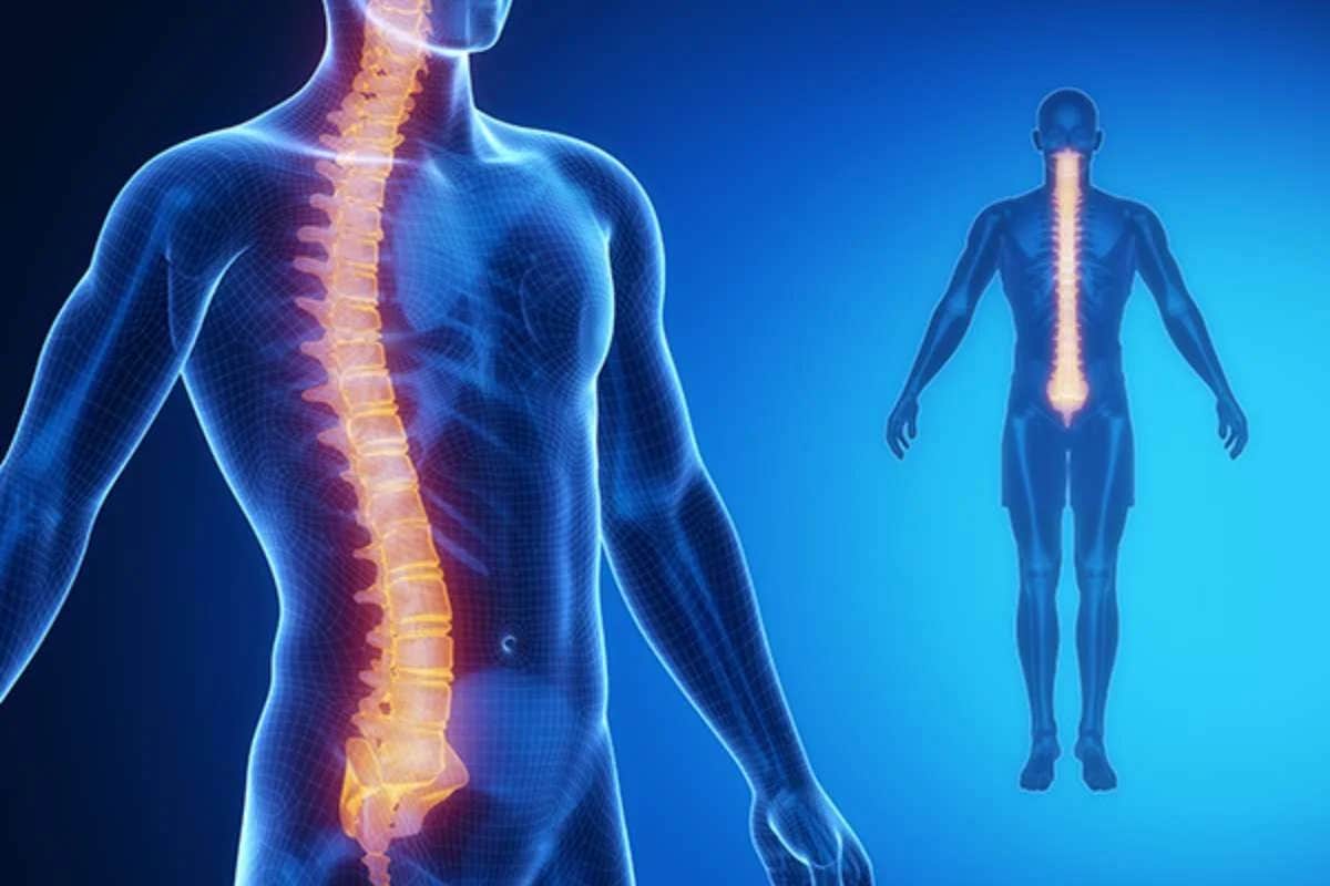
A single protein helps tiny blood vessel cells repair spinal cords and restore nerve function in new study. (CREDIT: iStock)
In the center of your body’s repair system are tiny cells with mighty power—pericytes. These small but active cells surround the smallest blood vessels and help maintain and heal tissues. Now, new research shows that pericytes may also unlock a future where damaged spinal cords can recover, not only structurally but also in function. For years, scientists questioned whether pericytes helped or harmed healing after spinal cord injuries. Now, there's a clearer answer—and it's hopeful.
From Problem to Possibility
After a spinal cord injury, many systems break down—not just the nerves. Blood vessels collapse, inflammation spreads, and normal cellular traffic halts. One of the first groups of cells to arrive at the site are pericytes. These cells migrate to the damaged area, but instead of helping, they sometimes become part of the problem. Scientists noticed that pericytes tightly grip sensory axons—the long, thread-like parts of nerve cells that send signals through the body. This contact disrupts the axons, making it harder for them to regrow.
But what if these same pericytes could be changed into helpers instead of hindrances?
Researchers from Ohio State University found a way to shift their role. By injecting a single dose of a protein called PDGF-BB (platelet-derived growth factor BB), they were able to reprogram the pericytes at the injury site. Once exposed to PDGF-BB, the cells reshaped themselves and started behaving differently. They stopped producing some molecules and began releasing others that encouraged axons to grow again.
“There's a lot more that can be learned and a lot that can be expanded,” said Andrea Tedeschi, senior study author and neuroscience professor. “But the more we worked on this, the more stunned we really were by the potency of this single treatment and how effective it was.”
Building Cellular Bridges
One of the most remarkable findings came from the way pericytes behaved after the PDGF-BB treatment. Normally, these cells crowd the lesion area but don’t build functional blood vessels or create a path for neurons to follow.
Related Stories
- Flexible electronic device wraps around the spinal cord - revolutionizing the treatment of spinal injuries
- Gel derived from cow mucus could revolutionize spinal cord treatment
- Breakthrough deep brain stimulation therapy restores walking after spinal cord injury
Once treated, however, pericytes changed shape—becoming long and slender—and started forming what researchers call “cellular bridges.” These bridges became physical pathways that supported axon growth.
In mouse studies, the researchers injected PDGF-BB directly into the spinal cord injury seven days after the injury—a delay similar to a nine-month healing window in humans. Four weeks later, axons had grown significantly more in treated mice than in those without the treatment. These axons appeared to use the pericyte-made bridges to escape the injury site.
“When we looked at formation of these pericyte structures that crossed the injury site, we saw the treatment promoted the growth of these bridges,” said first study author Wenjing Sun. “Most if not all of these regenerating axons were able to escape the injury site by riding these cellular bridges.”
Not only did these structures help axons grow, but the mice also regained movement in their back legs. Electrical tests showed that signals could now travel through the damaged area. The treated animals responded better to movement tests and showed fewer signs of nerve pain, a common and hard-to-treat result of spinal cord injuries.
Healing More Than Nerves
The power of this treatment went beyond just helping nerves grow. The protein PDGF-BB also helped rebuild tiny blood vessels. This is important because when blood flow stops after an injury, oxygen and nutrients can’t reach the cells trying to repair the damage. By restoring blood vessels, the researchers helped create a better environment for healing.
Further analysis showed that the pericytes didn’t change into another type of cell after treatment. They stayed true to their original form but took on new duties. While some genes linked to traditional pericyte behavior became less active, others that promoted healing turned on.
“There was a decrease in some classical pericyte markers, but a gain of some additional function linked to the attempt to rebuild cellular bridges and functional vessels,” Sun said. “From the overall gene signature in our data, they’re still classified as a pericyte.”
The treated area also had less inflammation, which usually causes more damage after spinal cord injuries. This added benefit likely helped reduce pain in the mice and allowed better long-term recovery.
From Mouse to Human Potential
Although this research began in mice, the team took steps to test its relevance in humans. In lab experiments, they used human pericytes and exposed them to PDGF-BB, then placed mouse neurons on top. The result? Just like in the mouse models, the axons started growing rapidly.
“To extend the clinical relevance of our findings, we cultured mouse neurons on top of human pericytes that were exposed to PDGF-BB,” said Tedeschi. “That was sufficient to trigger a growth-promoting effect, suggesting that this might really be a generalized phenomenon that is not restricted to mice.”
This opens the door for future treatments not only for spinal cord injuries but also for brain injuries, strokes, and diseases where nerve growth is slowed or stopped. Since PDGF-BB affects the environment around the injury and not just the neurons themselves, it may be possible to use it alongside other drugs that target nerve cells directly.
One such drug is gabapentin, which Tedeschi and Sun have studied before. It works by enhancing the growth ability of neurons. Combining it with PDGF-BB, which makes the injury site more friendly for healing, could offer a powerful two-part therapy. “We could combine both—modulating intrinsic properties of adult neurons with a drug, and what we are doing here, modulating the non-neuronal environment,” said Sun.
The Road Ahead
While these results are promising, several questions remain. Scientists want to find the best timing for delivering PDGF-BB after an injury. They also hope to learn the ideal dosage and whether a slow-release version would work better. Since pericytes take time to reach the injury site, there may be a window where the treatment is most effective.
Importantly, the treatment only needed to be given once to show powerful results in mice. But for people, a longer-lasting or repeated treatment might be needed. More studies will explore these details. What’s clear for now is that this single protein has the power to transform cells that once blocked healing into partners in repair. By changing how pericytes behave, scientists have found a way to rebuild the very structure of a damaged spinal cord.
For now, PDGF-BB shines as a surprising hero in the long fight to reverse the damage from spinal cord injuries—one cellular bridge at a time.
Research findings are available online in the journal Molecular Therapy.
Note: The article above provided above by The Brighter Side of News.
Like these kind of feel good stories? Get The Brighter Side of News' newsletter.