Breakthrough retina cell discovery could help treat blindness
Scientists have unveiled a groundbreaking use of nanotechnology to combat one of the most common causes of blindness in the world.
[July 31, 2023: Staff Writer, The Brighter Side of News]
Scientists have unveiled a groundbreaking use of nanotechnology to combat one of the most common causes of blindness in the world. (CREDIT: Creative Commons)
In an innovative leap forward in medical technology, scientists have unveiled a groundbreaking use of nanotechnology to combat one of the most common causes of blindness in the world. This marvel? A 3D ‘scaffold’ designed for the growth of retinal cells.
Led by Professor Barbara Pierscionek of Anglia Ruskin University (ARU), a determined team of researchers has successfully developed and grown retinal pigment epithelial (RPE) cells that remain vigorous and healthy for an astonishing duration of up to 150 days. These RPE cells are crucial elements of our visual system. Positioned just outside the neural segment of the retina, any damage to these cells can lead to significant vision degradation.
For the uninitiated, the revolutionary technology behind this breakthrough is named ‘electrospinning’. This marks the maiden instance of using electrospinning to craft a scaffold robust enough for RPE cells to grow upon. The implications of this discovery are staggering; it could fundamentally transform the treatment approach for age-related macular degeneration (AMD) - a predominant vision affliction globally.
Delving into the specifics, when this scaffold undergoes treatment with a steroid known as fluocinolone acetonide, renowned for its protective properties against inflammation, a remarkable phenomenon unfolds: the resilience of the eye cells skyrockets, fostering their growth. This revelation plays a monumental role in the evolution of ocular tissue development, primed for future transplantation into a patient's eye.
Related Stories
Shining a light on AMD, it has long been identified as a primary culprit behind blindness, especially in developed nations. With the ticking clock and the inescapable march of an ageing demographic, the specter of AMD looms even larger. Recent studies forecast a worrying trend: come 2050, Europe alone could witness an alarming 77 million individuals grappling with varying forms of AMD.
Tracing the origins of AMD leads us to the changes in Bruch’s membrane, a vital support structure for RPE cells, coupled with the breakdown of the choriocapillaris. This rich vascular bed is closely aligned with the other side of the Bruch’s membrane.
In most Western demographics, the primary trigger behind deteriorating sight remains the build-up of lipid deposits, known scientifically as drusen, which leads to the subsequent decay of parts of the RPE, the choriocapillaris, and outer retina. Contrarily, in developing regions, the typical manifestation of AMD stems from erratic blood vessel growth in the choroid. These abnormal vessels permeate into the RPE cells, catalyzing hemorrhaging, RPE or retinal detachment, and scarring.
Graphical study abstract. (CREDIT: Science Direct)
Given these challenges, the focus on RPE cells’ replacement surfaces as a beacon of hope, emerging as one of the most potent therapeutic avenues to address dire visual ailments like AMD. Researchers globally have pooled their resources, dedicating themselves to devise the most efficient methodologies to transplant these invaluable cells into the eye.
In an exclusive statement, lead author Professor Barbara Pierscionek, who also serves as the Deputy Dean (Research and Innovation) at ARU, elucidated, “This research has ushered in a paradigm shift, proving that nanofibre scaffolds, when paired with the anti-inflammatory agent fluocinolone acetonide, can dramatically enhance the growth, differentiation, and performance of RPE cells."
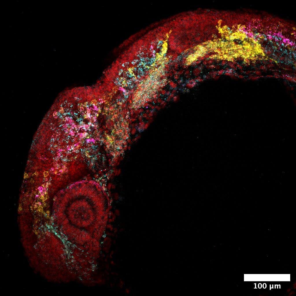
Genotoxic effects of FA-treated ENS on ARPE-19 cells. Cells were cultured on ENS or TCP for 5 days, prior to alkaline COMET assay to detect any DNA damage. (CREDIT: Science Direct)
Highlighting the evolutionary trajectory, she continued, "Traditionally, cells were cultivated on flat terrains, a practice that often yielded non-biologically relevant results. But with these avant-garde techniques, we've observed cells thriving within the three-dimensional realm afforded by these scaffolds."
Further expounding on its broader implications, Professor Pierscionek declared, "This system brims with potential. It could serve as a surrogate for Bruch’s membrane, offering a synthetic, non-toxic, and enduring base for the transplantation of retinal pigment epithelial cells. Recognizing pathological alterations in this membrane as a causative agent for ocular diseases like AMD, this groundbreaking innovation could be a panacea for millions on a global scale.”
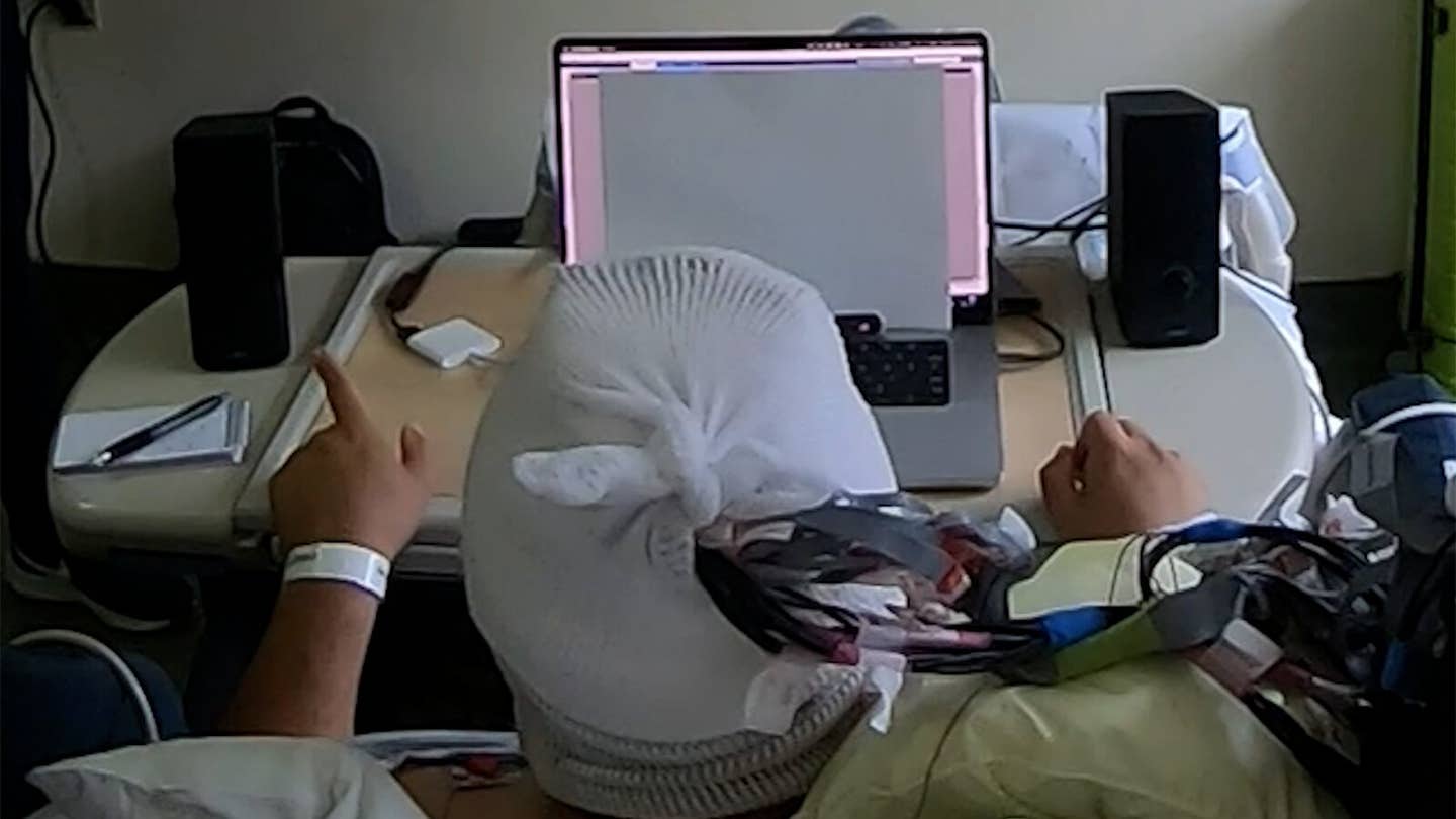
Representative images of unstained and fluorescently stained live ARPE-19 cells in culture with FA- treated ENS. Shown are unlabelled cells observed 5 minutes after seeding (A), labelled cells observed 30 minutes after seeding (B). Using confocal microscopy, cells infiltrated the PAN/Jeff series, whilst cells on PAN remained on the surface (C). Panel (D) shows SEM images of ARPE-19 cells cultured 24 h on FA- treated ENS. (CREDIT: Science Direct)
As the realm of nanotechnology fuses with medical science, we stand on the cusp of revolutionary treatments that could rewrite the narrative on eye health, providing hope and vision to countless souls worldwide.
Keywords: nanotechnology, 3D scaffold, retinal cells, Professor Barbara Pierscionek, Anglia Ruskin University, electrospinning, age-related macular degeneration, fluocinolone acetonide, ocular tissue development, Bruch’s membrane, RPE cells, choriocapillaris, vision deterioration, transplantation, breakthrough, AMD treatment.
Note: Materials provided above by The Brighter Side of News. Content may be edited for style and length.
Like these kind of feel good stories? Get the Brighter Side of News' newsletter.