Caltech scientists discover a ‘spooky’ way to double the resolution of light microscopes
Quantum entanglement, a phenomenon once described by Albert Einstein as “spooky action at a distance,” has been harnessed by scientists.
[May 3, 2023: Staff Writer, The Brighter Side of News]
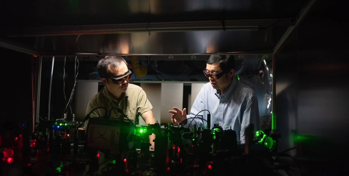
Zhe He (Postdoctoral Scholar Research Associate in Medical Engineering Andrew and Peggy Cherng Dept of Medical Engineering) and Lihong Wang (Bren Professor of Medical Engineering and Electrical Engineering). (CREDIT: Caltech)
Quantum entanglement, a phenomenon once described by Albert Einstein as "spooky action at a distance," has been harnessed by Caltech researchers to double the resolution of light microscopes, as reported in a recent paper published in the journal Nature Communications.
The team, led by Lihong Wang, Bren Professor of Medical Engineering and Electrical Engineering, has developed a groundbreaking microscopy technique called quantum microscopy by coincidence (QMC), which takes advantage of quantum entanglement to improve microscope resolution without damaging the specimens being observed.
Quantum entanglement occurs when two particles become linked in such a way that the state of one particle is tied to the state of the other, regardless of their distance from each other. In Wang's QMC technique, the entangled particles are photons.
When two entangled photons, also known as a biphoton, behave as a single particle with twice the momentum of a single photon, they have a wavelength half that of individual photons. This is crucial to the QMC technique, as reducing the wavelength of light used in microscopy enables the observation of smaller features, resulting in increased resolution.
Related Stories
Traditionally, using light with shorter wavelengths to achieve higher resolution has been challenging due to the higher energy carried by shorter wavelengths. This can damage delicate specimens, such as living cells. However, QMC overcomes this limitation by utilizing biphotons that have the lower energy of longer-wavelength photons while maintaining the shorter wavelength of higher-energy photons.
Wang explains, “Cells don’t like UV light. But if we can use 400-nanometer light to image the cell and achieve the effect of 200-nm light, which is UV, the cells will be happy, and we’re getting the resolution of UV.”
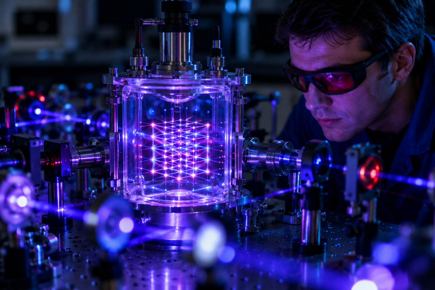
To achieve this, the Caltech team constructed an optical apparatus that converts laser light into biphotons using a special crystal. Despite the rarity of this conversion, occurring in only one in a million photons, the researchers were able to separate the biphoton into two discrete photons, each taking a different path.
The quantum microscopy apparatus. (CREDIT: Lance Hayashida/Caltech)
One photon passes through the object being imaged, while the other does not. These paired photons, known as the signal and idler photons, remain entangled as a biphoton with half the wavelength, despite the presence of the object and their separate pathways. A detector connected to a computer then builds an image of the cell based on the information carried by the signal photon.
Wang's lab was the first to create a viable system using biphoton imaging. “We developed what we believe a rigorous theory as well as a faster and more accurate entanglement-measurement method. We reached microscopic resolution and imaged cells,” Wang stated.
A diagram of the quantum microscopy apparatus. CW continuous wave, GL Glan-Laser polarizer, HWP half-wave plate, VWP variable wave plate, BBO β-barium borate crystal, BPF 532 nm bandpass filter, PBS polarizing beam splitter, EMCCD electron multiplying charge-coupled device camera. f0 = 50 mm, f1 = 180 mm, f2 = 9 mm, f3 = 300 mm, and f4 = 200 mm. The source Fourier plane P0 is set at the Fourier plane of the BBO crystal. (CREDIT: Caltech)
While there is no theoretical limit to the number of photons that can be entangled, each additional entangled photon further increases the momentum of the resulting multiphoton and decreases its wavelength. Wang notes that future research could explore the entanglement of even more photons, but cautions that each additional photon would further reduce the already low probability of successful entanglement.
This breakthrough in microscopy utilizing quantum entanglement opens new possibilities for imaging delicate biological specimens without damaging them, promising exciting advancements in the field of microscopy and our understanding of the microscopic world.
Side by side comparison of microscope resolution. (CREDIT: Caltech)
Paper co-authors are Zhe He and Yide Zhang, both postdoctoral scholar research associates in medical engineering; medical engineering graduate student Xin Tong (MS ’21); and Lei Li (PhD ’19), formerly a medical engineering postdoctoral scholar and now an assistant professor of electrical and computer engineering at Rice University.
Funding for the research was provided by the Chan Zuckerberg Initiative and the National Institutes of Health.
For more science and technology news stories check out our New Innovations section at The Brighter Side of News.
Note: Materials provided above by The Brighter Side of News. Content may be edited for style and length.
Like these kind of feel good stories? Get the Brighter Side of News' newsletter.