Cutting-edge AI technology reinvents surgery through real-time diagnoses
When a patient goes into surgery to remove a tumor or address a disease, often the precise course of action is not pre-determined.
[Mar. 22, 2023: Aaron Sanborn, Brigham and Women's Hospital]
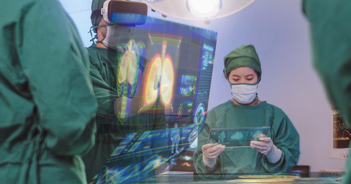
When surgeons send samples to a pathologist for examination, both speed and accuracy are of the essence. (CREDIT: Creative Commons)
When a patient goes under the knife for a surgical procedure to remove a tumor or address a disease, the precise course of action is often not pre-determined. In order to determine the extent of the tissue that needs to be excised, surgeons need to possess a thorough understanding of the ailment they are dealing with, including factors such as the tumor's margins, stage, and whether it is benign or malignant. These determinations often rely on the collection, analysis, and diagnosis of a disease while the patient is still under anesthesia on the operating table.
Once surgeons collect samples, they send them to a pathologist for swift and accurate examination. While a gold-standard approach is available, it is not always practical as it can take an extended period of time to examine tissues, which is not ideal when the patient is still in surgery. A faster method involves freezing the tissue, which can introduce artifacts that may complicate diagnosis.
Researchers from the Mahmood Lab at Brigham and Women's Hospital, a member of the Mass General Brigham healthcare system, in collaboration with Bogazici University, have discovered an innovative method that employs artificial intelligence to bridge the gap between frozen sections and the gold-standard approach. This method improves image quality, thereby increasing the accuracy of rapid diagnostics.
Findings are published in Nature Biomedical Engineering.
Related Stories
“We are using the power of artificial intelligence to address an age-old problem at the intersection of surgery and pathology,” said corresponding author Faisal Mahmood, PhD, of the Division of Computational Pathology at BWH. “Making a rapid diagnosis from frozen tissue samples is challenging and requires specialized training, but this kind of diagnosis is a critical step in caring for patients during surgery.”
When it comes to providing final diagnoses, pathologists rely on formalin-fixed and paraffin-embedded (FFPE) tissue samples. Although this method produces high-quality images, the process is often laborious and can take anywhere from 12 to 48 hours.
For a rapid diagnosis, pathologists employ cryosectioning, a quick diagnostic method, that involves rapidly freezing tissue, cutting sections, and examining them under a microscope. Although this method takes only minutes, it can deform cellular features and damage fragile tissue.
Cryosection to FFPE translation workflow. (CREDIT: Nature Biomedical Engineering)
A deep-learning model developed by Mahmood and collaborators can convert between frozen sections and the more commonly used FFPE tissue.
The team showed in their publication that the method was applicable for subtyping various cancer types, such as glioma and non-small-cell lung cancer. To confirm their results, they conducted a reader study in which pathologists were recruited to diagnose images that had been processed using both the AI method and traditional cryosectioning images.
AI-FFPE method architecture. (CREDIT: Nature Biomedical Engineering)
Not only did the AI method enhance image quality, but it also increased the diagnostic precision among specialists. Additionally, the algorithm underwent testing using independently collected data from Turkey.
The authors suggest that future clinical studies should be conducted to confirm the efficacy of the AI method and its ability to enhance diagnostic accuracy and surgical decision-making in real hospital settings.
According to Mahmood, "Our findings demonstrate that AI has the potential to simplify and make time-sensitive diagnoses more accessible to pathologists. This technology can be applied to various types of cancer surgery, presenting numerous opportunities for enhancing patient care and diagnosis."
Note: Materials provided above by Brigham and Women's Hospital. Content may be edited for style and length.
Like these kind of feel good stories? Get the Brighter Side of News' newsletter.