First-ever portable, radiation-free, scanner can peer deep inside the human body
A cutting-edge technology named Magnetic Particle Imaging (MPI) offers a radiation-free option for in-depth insights.
[July 27, 2023: Staff Writer, The Brighter Side of News]
A cutting-edge technology named Magnetic Particle Imaging (MPI) promises not only to revolutionize the field of medical imaging but also offers a radiation-free option for in-depth insights. (CREDIT: Nature Scientific Reports)
In today's fast-paced medical landscape, the invaluable role of imaging technologies is undeniable. Tools such as computed tomography (CT), magnetic resonance imaging (MRI), positron emission tomography (PET), and ultrasound are indispensable players in the diagnostics arena.
Each presents a unique lens through which to view the intricate inner workings of the human body, allowing physicians to detect abnormalities or assess functional processes.
Now, a promising addition is making its mark on this list. A cutting-edge technology named Magnetic Particle Imaging (MPI) promises not only to revolutionize the field of medical imaging but also offers a radiation-free option for in-depth insights.
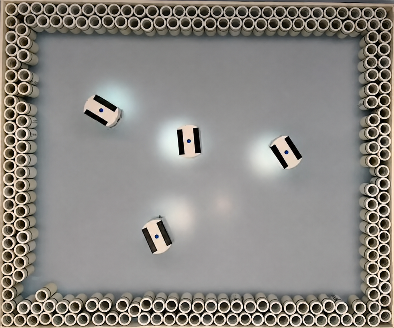
Developed by a formidable team of physicists and medical professionals at Julius-Maximilians-Universität Würzburg (JMU), this new technology utilizes a portable scanner to capture dynamic processes inside the human body - imagine visualizing blood flow in real-time.
Related Stories
Spearheading this groundbreaking study were Professor Volker Behr and Dr. Patrick Vogel, both representing the University's Institute of Physics. Their findings, which could chart a new course for the future of medical imaging, were recently published in the esteemed journal Nature Scientific Reports.
The uniqueness of MPI lies in its use of magnetic nanoparticles as markers. Volker Behr notes, "As with positron emission tomography, which relies on the administration of radioactive substances as markers, this method has the great advantage of being sensitive and fast without 'seeing' interfering background signals from tissue or bone."
Unlike PET, which detects gamma rays from a radioactive marker, MPI's focus is on the response signal from the magnetic nanoparticles when they're subjected to changing magnetic fields.
The iMPI scanner is so small and light that you can take it with you and use it almost anywhere. This is a first important step towards radiation-free intervention. (CREDIT: Patrick Vogel / Stefan Herz)
Dr. Patrick Vogel, the study's primary author, clarifies, "In this process, the magnetisation of nanoparticles is specifically manipulated with the help of external magnetic fields, allowing us not only to detect their presence but also to ascertain their spatial position in the human body."
The Evolution of MPI
MPI isn't a novel idea. Back in 2005, Philips demonstrated the potential of this technology, but the samples were restricted to a mere few centimetres. Efforts to adapt MPI for human-sized scans led to hefty, unwieldy, and expensive equipment.
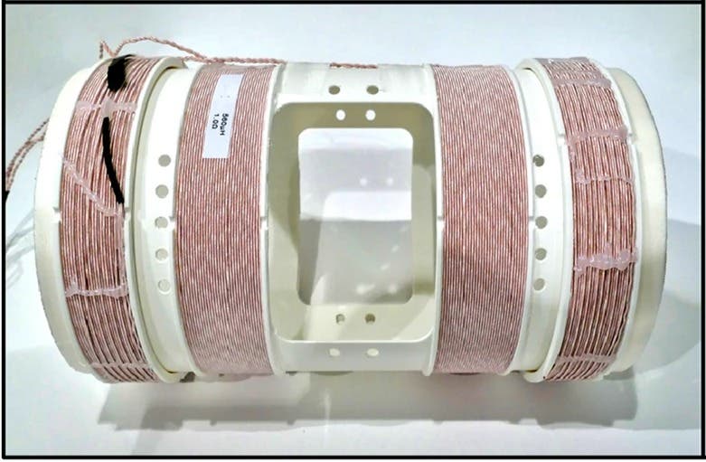
Sketch of the concept for compact human-sized MPI scanners using a field-free line (FFL). (a,b) Two pairs of saddle-coils are driven with a frequency f1 = f2 = 60 Hz and a phase shift of 90 degrees resulting in a traveling FFL along the symmetry axis of the scanner. (c) Two additional solenoid coils in Helmholtz configuration (CH3) are used for FFL displacement within the X–Y-plane. (d) Running CH3 with a frequency f3 ≫ f1, the FFL is steered along a sinusoidal trajectory through the FOV generating 2D projections. (CREDIT: Nature Scientific Reports)
Fast-forward to 2018, the team under the visionary guidance of Professor Behr and Dr. Vogel pioneered an innovative approach to create complex magnetic fields in a compact form. This was made possible thanks to the generous support from the German Research Foundation (DFG). After years of relentless research, the result was a specially designed MPI scanner, coined as the interventional Magnetic Particle Imaging (iMPI).
Dr. Vogel proudly states, "Our iMPI scanner is so small and light that you can take it almost anywhere." Its capabilities were further highlighted in a simultaneous real-time comparison with a specialized X-ray device, commonly used in university hospitals for angiography.
Two different approaches for receive coils are demonstrated: left: the rigid-body receive coils with different diameters (dblack = 11 cm, dwhite = 18 cm) provide four coils each to be used as gradiometer. Both provide a dedicated X-ray window. Right: a flexible receive coil consisting of two racetrack coils assembled on a flexible material. (CREDIT: Nature Scientific Reports)
The proficiency of the iMPI scanner was put to the test on a realistic vascular phantom by Professor Thorsten Bley and Dr. Stefan Herz's team from the Interventional Radiology Department at Würzburg University Hospital. Dr. Herz, reflecting on the device's potential, remarked, "This is a first important step towards radiation-free intervention. MPI has the potential to change this field for good."
With the initial success of the iMPI device, Behr and Vogel are not resting on their laurels. Their quest is focused on refining the scanner to achieve even better image quality.
As we navigate the complex domain of medical imaging, the arrival of tools like MPI offers not just another technological marvel but a beacon of hope for safer, more efficient diagnostic procedures. As researchers and medical practitioners continue to push the boundaries of what's possible, it is clear that the future of medical imaging, with MPI at the helm, looks promising and radiation-free.
For more science stories check out our New Innovations section at The Brighter Side of News.
Note: Materials provided above by The Brighter Side of News. Content may be edited for style and length.
Like these kind of feel good stories? Get the Brighter Side of News' newsletter.