Healing power of light: New treatment offers hope for millions suffering from corneal diseases
A groundbreaking new study by uOttawa researchers presents a new hope for millions suffering from corneal diseases worldwide.
[Sept 1, 2023: Staff Writer, The Brighter Side of News]
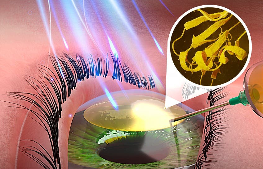
The study discovered a biomimetic material, which when pulsed with low-energy blue light, can potentially reshape and fortify damaged corneas. (CREDIT: Alarcon)
A groundbreaking new study by uOttawa researchers and their collaborators presents a new hope for millions suffering from corneal diseases worldwide.
The study discovered a biomimetic material, which when pulsed with low-energy blue light, can potentially reshape and fortify damaged corneas. This innovative finding is set to be a game-changer, with potential to bring relief to millions affected by vision impairment due to thinning corneas.
Nature-inspired Innovation
This revolutionary approach to corneal repair is based on biomimetic design, a method of innovating by drawing inspiration from nature. The multidisciplinary research team from uOttawa has unveiled compelling results, establishing that this newly developed light-activated material could be instrumental in reshaping and reinforcing damaged corneal tissue. This, in turn, can greatly enhance the healing and recovery process of the cornea, offering a new lifeline for many.
Corneal diseases impact tens of millions of people globally. The current predominant treatment is corneal transplantation, which has its limitations. Many conditions, like keratoconus, can lead to vision loss and, unfortunately, only a fraction of patients are eligible for transplants.
Related News
Dr. Emilio Alarcon, an Associate Professor at the uOttawa Faculty of Medicine, expresses high hopes for the new technology: “Our technology is a leap in the field of corneal repair. We are confident this could become a practical solution to treat patients living with diseases that negatively impact corneal shape and geometry, including keratoconus.”
The cornea plays an indispensable role in our vision, serving as the protective dome-like structure of the eye, controlling and directing light rays. Injuries or infections can result in the cornea's scarring, which impairs vision.
The Pioneering Technology
Published in the esteemed scientific journal, Advanced Functional Materials, the team’s research revolves around biomaterials consisting of short peptides and natural polymers called glycosaminoglycans.
Schematic research strategy followed in this work. Top: Steps 1 and 2: Development of a low dosage blue-light activated material for in situ corneal repair using collagen-like peptides (CLPs) to replace gelatin, the initial test polymer. Step 3 depicts the creation of a library for the different compositions of the formulations. Pulses of blue light were applied in steps 2, 6 and 7. Bottom: Custom-designed peptides with photoreactive moieties used in this study. (CREDIT: Advanced Functional Materials)
Once a minuscule pocket is surgically crafted, this viscous liquid is injected into the corneal tissue. Remarkably, when exposed to pulses of low-energy blue light, this injected hydrogel solidifies, transforming into a transparent, tissue-like 3D structure within mere minutes. As Dr. Alarcon notes, its properties closely mirror those of pig corneas.
Promising Test Results
The team's experiments, using a rat model, indicated that this light-activated hydrogel can fortify corneas without adverse effects. The research employed a significantly lesser dosage of blue light compared to other studies, and has been successfully tested in an ex vivo pig cornea model. Before it can progress to clinical human trials, further testing in larger animal models is essential.
Dr. Emilio Alarcon — Associate Professor, Faculty of Medicine and researcher at University of Ottawa Heart Institute. (CREDIT: UOttawa)
Highlighting the material's efficiency, Dr. Alarcon commented: “Our material was engineered to harness blue light energy, facilitating the immediate assembly of the material into a cornea-like structure. Preliminary data suggests that the materials are non-toxic and can remain stable for several weeks in an animal model. We project similar non-toxic stability in human corneas.”
Decade-long Development
A monumental undertaking, the meticulous research took over seven years to materialize to its current published form.
Using GelMa to identify the optimal composition for a peptide-based light-activated cornea filler. Schematic depiction for the main steps used in the preparation of the light activated materials. (CREDIT: Advanced Functional Materials)
Recounting the rigorous process, Dr. Alarcon mentions, “Each component of the technology, from the light source to the molecules, was engineered from scratch. The goal was to ensure the technology would be clinically translatable, aligning with stringent sterility standards.”
A Look Ahead
With patent negotiations underway, this promising discovery could soon pave the way for groundbreaking treatments in the world of ophthalmology. Spearheading the material design aspect of the research, Dr. Alarcon collaborated with prominent figures, including Dr. Marcelo Muñoz and Aidan MacAdam from uOttawa.
Notable interdisciplinary collaborators were Dr. May Griffith and Dr. Isabelle Brunette from Université de Montréal, experts in cornea regeneration and corneal transplant, respectively.
For the millions grappling with deteriorating vision and the agonizing wait for corneal transplants, this innovative approach promises not only improved sight but also a brighter future.
Note: Materials provided above by The Brighter Side of News. Content may be edited for style and length.
Like these kind of feel good stories? Get the Brighter Side of News' newsletter.