MIT scientists develop wearable ultrasound scanner to detect breast cancer earlier
MIT researchers designed a wearable ultrasound device that could allow women to detect tumors when they are still in early stages.
[July 29, 2023: Staff Writer, The Brighter Side of News]
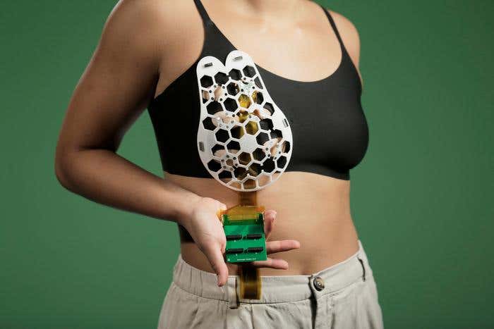
In hopes of improving the survival rate for breast cancer patients, MIT researchers designed a wearable ultrasound device that could allow women to detect tumors when they are still in early stages. (CREDIT: Canan Dagdeviren)
In oncology, the chances of survival can be dramatically influenced by the time of diagnosis. For instance, with breast cancer, if caught in its nascent stages, the survival rate approaches an astounding 100 percent. Conversely, that rate plummets to a bleak 25 percent if the cancer is detected in advanced stages. This chasm in prognosis is primarily due to the nature of the disease progression and the methods available for early detection.
In a groundbreaking study led by the Massachusetts Institute of Technology (MIT), researchers have unveiled a wearable ultrasound device designed to empower individuals with the ability to detect potential breast tumors in their earliest stages. The device is particularly beneficial for patients at high risk of developing breast cancer who are often at the mercy of the interval between routine mammograms.
At the core of this ingenious invention is a flexible patch capable of adhering to a bra. This non-invasive device equips the wearer to move an ultrasound tracker across the patch, enabling them to image breast tissue from various angles. In tests conducted by the team, the ultrasound images derived from the device were found to be of resolution comparable to that obtained by the ultrasound probes typically used in medical imaging centers.
“We changed the form factor of the ultrasound technology so that it can be used in your home. It’s portable and easy to use, and provides real-time, user-friendly monitoring of breast tissue,” explains Canan Dagdeviren, an associate professor in MIT’s Media Lab and the senior author of the study.
Related Stories:
The research team comprises a diverse pool of talent, including MIT graduate student Wenya Du, Research Scientist Lin Zhang, Emma Suh ’23, and Dabin Lin, a professor at Xi’an Technological University. Their pioneering work has been published in the prestigious journal Science Advances.
The development of the wearable ultrasound device stemmed from a deeply personal experience for Dagdeviren. Her late aunt, Fatma Caliskanoglu, was diagnosed with late-stage breast cancer at the age of 49 despite regular screenings. Unfortunately, she passed away six months later. This tragic incident, witnessed firsthand by Dagdeviren, motivated her to conceptualize a diagnostic device that could be integrated into everyday clothing, thereby promoting more frequent screenings for high-risk women.
Cancers that appear between regular mammogram screenings — termed interval cancers — constitute 20 to 30 percent of all breast cancer cases. These tend to be more aggressive than those found during routine screenings. Dagdeviren’s vision targets this issue head-on, as she remarks, “My goal is to target the people who are most likely to develop interval cancer. With more frequent screening, our goal to increase the survival rate to up to 98 percent.”
The overview of the design of the cUSBr-Patch. Schematic of a cUSBr-Patch on the body. Exploded view of the cUSBr-Patch to illustrate its four main components: a soft fabric bra to serve as a familiar intermediary layer, a honeycomb patch as the outside layer to provide structure and guidance of the 1D array, the tracker to hold and rotate the 1D array, and the single crystal–based 1D phased array. (CREDIT: Science Advances)
In the pursuit of this vision, Dagdeviren designed a miniaturized ultrasound scanner that encourages user-initiated imaging at any time. Built on the same ultrasound technology used in medical imaging centers, the device incorporates a novel piezoelectric material that facilitated the miniaturization of the ultrasound scanner.
To transform the scanner into a wearable, the researchers developed a flexible, 3D-printed patch with honeycomb-like openings. This patch can be magnetically attached to a bra, with the openings allowing the ultrasound scanner to make contact with the skin. The scanner, contained within a small tracker, can be maneuvered to six different positions for comprehensive breast imaging. The scanner can also be rotated to capture images from various angles, and it does not necessitate specialized expertise for operation.
The structure and properties of the Yb/Bi-PIN-PMN-PT single crystal. Phase diagram of the PIN-PMN-PT single crystal. Photograph of the as-grown Yb/Bi-PIN-PMN-PT single-crystal boule and polished cross section. Five samples, I to V, were diced from different positions of the rod for routine characterization. (CREDIT: Science Advances)
Anantha Chandrakasan, dean of MIT’s School of Engineering, the Vannevar Bush Professor of Electrical Engineering and Computer Science, and one of the authors of the study, speaks highly of the technology, stating, “This technology provides a fundamental capability in the detection and early diagnosis of breast cancer, which is key to a positive outcome. This work will significantly advance ultrasound research and medical device designs, leveraging advances in materials, low-power circuits, AI algorithms, and biomedical systems.”
The team tested their device on a 71-year-old woman with a history of breast cysts at the MIT Center for Clinical and Translational Research. The device successfully detected cysts as small as 0.3 centimeters in diameter — the size of early-stage tumors. They also confirmed that the device achieved resolution comparable to traditional ultrasound, capable of imaging tissue up to 8 centimeters deep.
Catherine Ricciardi, nurse director at MIT’s Center for Clinical and Translational Research and an author of the study, champions the technology’s potential, commenting, “Access to quality and affordable health care is essential for early detection and diagnosis. As a nurse I have witnessed the negative outcomes of a delayed diagnosis. This technology holds the promise of breaking down the many barriers for early breast cancer detection by providing a more reliable, comfortable, and less intimidating diagnostic.”
For the device to relay ultrasound images, the scanner must currently connect to the same kind of ultrasound machine used in imaging centers. However, the researchers are developing a miniaturized version of the imaging system approximately the size of a smartphone.
The wearable ultrasound patch is reusable and could become a common home-based device for individuals at high risk for breast cancer. Its potential for increasing accessibility is profound, especially for those who do not have regular access to screening.
The piezoelectric and acoustic performance of 1D phased array. Electrical impedance spectrum of a single element of the phased array. Measured waveform and frequency spectrum of the single element of the phased array. (CREDIT: Science Advances)
Tolga Ozmen, a breast cancer surgeon at Massachusetts General Hospital and an author of the study, identifies the device as a significant development, stating, “Breast cancer is the most common cancer among women, and it is treatable when detected early. One of the main obstacles in imaging and early detection is the commute that the women have to make to an imaging center. This conformable ultrasound patch is a highly promising technology as it eliminates the need for women to travel to an imaging center.”
Looking ahead, the researchers aim to leverage artificial intelligence to scrutinize how the images evolve over time, potentially providing more accurate diagnostics than relying on the assessment of a radiologist comparing images taken years apart. The team also plans to explore the possibility of adapting the ultrasound technology to scan other parts of the body.
In essence, MIT’s wearable ultrasound device marks a significant leap forward in the democratization of healthcare and the battle against breast cancer. It encapsulates the possibility of a future where access to medical diagnostics isn’t limited by geography or time constraints, and instead, places the power of early detection directly into the hands of those who need it the most.
For more science stories check out our New Innovations section at The Brighter Side of News.
Note: Materials provided above by The Brighter Side of News. Content may be edited for style and length.
Like these kind of feel good stories? Get the Brighter Side of News' newsletter.