New AI tool can detect early signs of aging from routine chest X-rays
AI analysis of chest X-rays may reveal early signs of aging and disease risk better than DNA-based aging clocks.

 Edited By: Joseph Shavit
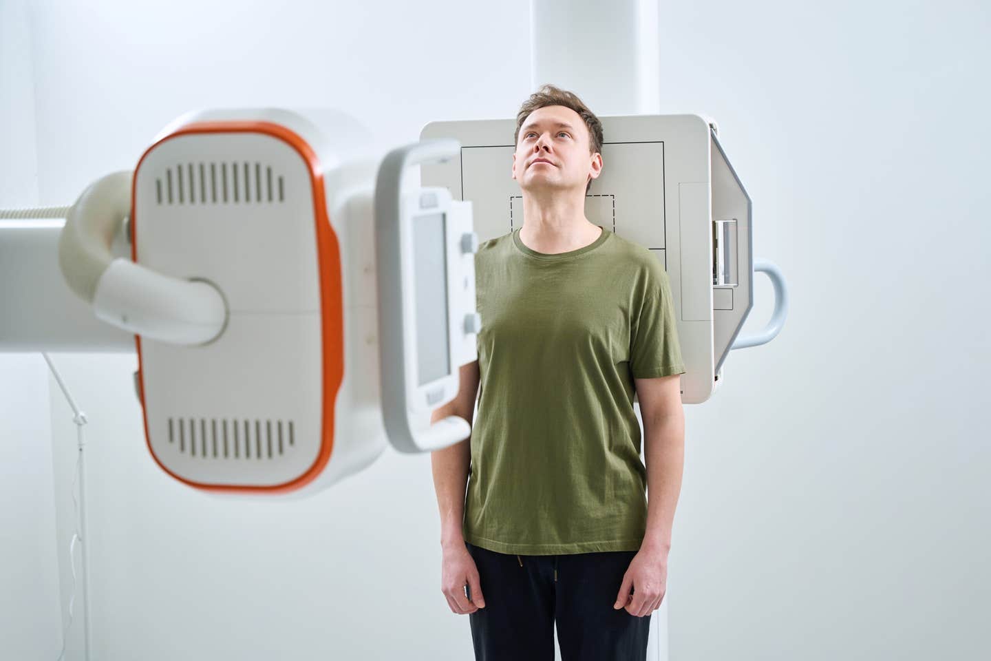
A new study finds that artificial intelligence can estimate biological aging from routine chest X-rays. (CREDIT: Shutterstock)
Chronological age is a primary method for assessing healthcare managers' decisions regarding how to treat and care for patients. Chronological age does not account for other factors that affect the rate of an individual's aging process, such as genetics, lifestyle behaviors, medical history, environment, and diet.
In response to this limitation, scientists have developed "biological age clocks," which allow the assessment of an individual's biological age. The biological age of an individual may indicate earlier detection of diseases such as heart disease and lung disease. These chronologic and biological ages differ for many reasons: An individual may develop heart disease and lung disease much earlier than others of the same chronological age.
The most well-known biological age clocks are epigenetic biological age clocks. Epigenetic biological age clocks measure changes in chemical structure to determine an individual's biological age through measuring changes in DNA.
Original epigenetic biological age clocks, including the Horvath epigenetic clock, were developed to be closely correlated with chronological age. Newer epigenetic biological age clocks, including the DNAm PhenoAge clock, use other factors to determine risk for death, disease, and overall health.
Comparing Imaging and DNA Aging Clocks
CXR-Age uses the application of deep learning through the analysis of chest X-rays to measure biological age based upon the visible effects of aging on the heart and lungs. This assessment is based on the progression of changes to the shape and structure of the organs due to the aging process. The changes to the shape and structure of the organs will develop many years before symptoms are evident in these organs. Evaluating Differences between Imaging and Biological Ageing
One study examined data collected from 2,097 participants who had enrolled in the Project Baseline Health Study, a US-based project involving several locations over time. The average age of participants was just over 51 years and slightly more than half were female. Many of the participants completed either a chest x-ray or a DNAmeth opsy, or both.
For those with complete information, the average CXR-Age according to the CXR-Age model was approximately 53 years of age, compared to the average Horvath-Clock Age of approximately 55 years and the average DNAm PhenoAge of approximately 39 years, indicating that while both DNA-based models closely matched biological age, the more relevant question was whether they were capable of detecting the onset of diseases earlier. That is the basis of the value added by using imaging.
CXR-Age, Heart Disease Link
The most significant finding from this research study is related to Coronary Artery Calcium (CAC), which reflects plaque deposition in arterial walls and is a well-established risk factor for heart disease. Every year, the CXR-Age is greater than the predicted value, the CAC would increase by 10% and be positively associated with CAC according to DNAm PhenoAge. The Horvath Age Clock did not show a substantive relationship.
This relationship persisted even when restricting the analysis to individuals aged 45 and older. CXR-Age is also closely related to cardiovascular risk scores for 10 years, and every year a person has accelerated CXR-Age, the amount of risk associated with their heart would increase by 4% in each age category.
Through this research study, it is therefore proposed that CXR's provide a means by which an individual can be assessed for cardiovascular risk changes long before the presence of abnormalities in routine blood tests and evaluations.
The Relationship Between Age and Lung Function Declines
Using The CXR-Age model has demonstrated a clear link to lung health, with multiple measures, including the FEV1/FVC ratio and diffusion capacity (DLCO) being examined as well to determine the amount of oxygen transferred into the bloodstream as well as the amount of air displaced by the lungs (for every additional CXR-Age year, lung diffusion capacity decreased by 0.74%).
While the CXR-Age model identified a decline in the DLCO along with other measures of lung function (except for peak airflow), the connection found with DNAm PhenoAge has been less robust and only supported by the association to duration of the DLCO. The Horvath clock again did not exhibit any statistically significant correlation.
Most importantly, all CXR-Age relationships remained significant regardless of smoking status, as cigarette smokers and non-smokers exhibited similar levels of association, which indicates that the CXR-Age model has identified changes in the lungs related to aging, rather than changes caused solely by smoking damage.
Declines in Overall Health and Physical Functionality
While the CXR-Age model has been applied to examine organ systems, it has also been used to evaluate individual functions, including hand grip strength, walking speed, balance, number of repetitions of "sit-to-stand" tests, and distance walked in six minutes. These functional tests reflect frailty and an individual’s ability to perform daily living activities.
During the course of the study, the CXR-Age model has consistently demonstrated a greater association with deteriorating physical performance than with the DNAm PhenoAge or Horvath clock models. Every additional year of accelerated aging (CXR-Age) was associated with lower levels of grip strength, slower walking times, shorter durations of balance maintenance, and poorer results on sit-to-stand functional tests. Additionally, individuals with increased disability scores (based on the Sheehan disability scale and WHODAS 2.0) exhibited increased CXR-Age scores.
Along with showing stronger correlations with physical performance scores, the DNAm PhenoAge was associated with fewer and weaker connections to physical performance measures than the CXR-Age model, while the Horvath clock was associated with little or no connection to physical performance measures.
Clues from Blood Proteins and Imaging Review
This age-related forward-thinking model CXR-Age, was valid for predicting disease on chest X-rays at all ages, with the most notable effects seen in individuals aged 45 years and older. As such, this model may prove particularly worthwhile for identifying potential age-related health risks during middle-aged years.
Humans have long assumed their biological age could be measured through how much action or inaction a person has engaged in throughout life thus far. The research team also recognized this belief; to investigate the biological basis for this assumption, they analyzed plasma from 957 individuals and looked specifically at two proteins known to be critical to both inflammation and the aging process, CDH13 and ApoD.
This finding, in conjunction with the previous findings of this study (DNAm PhenoAge), indicates that the CXR-Age forward-thinking model potentially has common biological pathways through both DNAm PhenoAge and the proteins examined.
In addition to examining the link between CXR-Age and the two proteins, the team of researchers reported that they had radiologists review a series of X-rays ranging from very low to very high CXR-Age acceleration. The review of the X-rays from both directions highlighted some features observed on the lower CXR-Age X-ray series that consistently indicated clear lungs and normal sized hearts.
Conversely there were clear indicators of increased lung markings, thickened airways (indicating some form of respiratory disease), fluid build-up in the lungs, enlargement of the heart and/or lungs as observed by the radiologists in the CXR-Age X-rays with the scores. Therefore, these visible and abnormal features provided insight into how the CXR-Age forward-thinking model learns to identify these features.
Opinion of Research Experts
According to the new study published in Journal of Gerontology, it would be reasonable to suggest that artificial intelligence could be used to assess how quickly our bodies are aging based on subtle indications found in routine medical imaging.
"The results of this study suggest that deep learning applied to common medical imaging modalities will aid in determining how our body organs are aging. As the information provided from these studies progresses, we are hopeful these findings will help identify patients at risk of developing age-related disease before the onset of age-related symptoms, Douglas P. Kiel, MD, MPH, Director of the Musculoskeletal Research Center at the Marcus Institute for Aging Research and co-author of this study, told the Brighter Side of News.
"The AI tools developed in conjunction with CXR-Age hold great potential for enabling the deployment of cost-efficient methods of assessing and reducing disease risk associated with age before the onset of age-related illness," he continued.
Despite these limitations, the discovery is an indication that one day a routine chest X-ray could proactively identify emerging health risks.
Research findings are available online in the Journal of Gerontology.
Related Stories
- Scientists create color X-ray images of the future
- New AI model revolutionizes medical imaging with 90% less computing power
- AI predicts patient's race from x-ray images with 99% accuracy - is that as good thing?
Like these kind of feel good stories? Get The Brighter Side of News' newsletter.
Shy Cohen
Writer