New brain imaging system links light, sound, and metabolism
A new imaging system called label free multiphoton photoacoustic microscopy uses sound to track brain metabolism at single-cell resolution.
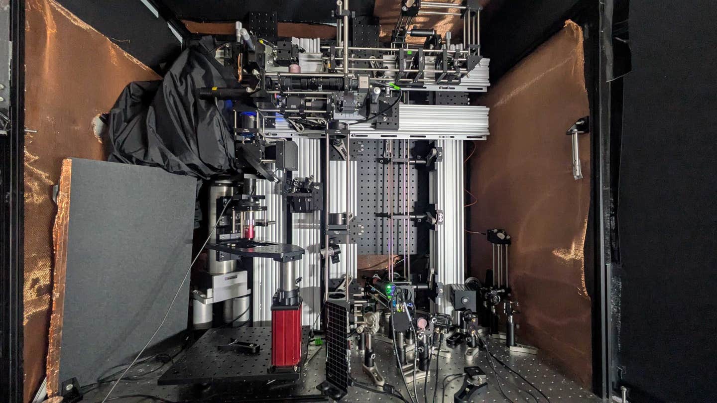
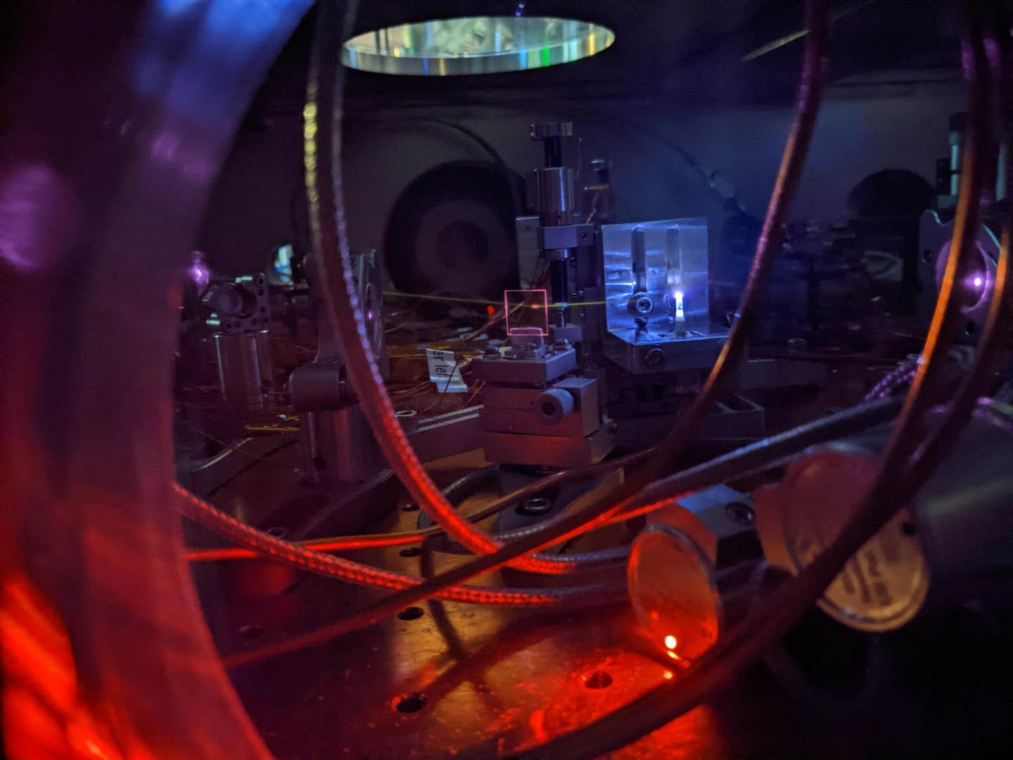
A picture of the microscope system. (CREDIT: Tatsuya Osaki)
For decades, scientists have worked to peer deeper into the brain. They have wanted to see not just surface activity in the cortex but also the hidden layers beneath, such as the hippocampus, where memory and learning take shape. A new breakthrough may finally open that window.
A team of researchers from MIT has built a microscope that listens to brain metabolism. Instead of relying only on light, this system uses bursts of sound to track the molecules that fuel brain activity. The innovation is known as label free multiphoton photoacoustic microscopy, and it could transform how we study the brain and diagnose disease.
Listening to Molecules Instead of Adding Labels
The technique is designed to detect a molecule called NADH, also known as NAD(P)H when in its related form. This small coenzyme is central to metabolism, the process that drives life inside every cell. When neurons fire or when disease disrupts brain chemistry, NADH levels change. That makes it a powerful biomarker for both healthy activity and disorders.
Traditional microscopes try to capture NADH using near-ultraviolet fluorescence. But there is a problem. In brain tissue, this fluorescence can only be seen about 100 microns deep—roughly the thickness of a human hair. That limit has long frustrated neuroscientists.
The new method avoids that wall by turning light into sound. It uses a near-infrared femtosecond laser—pulses lasting a quadrillionth of a second—to excite NADH in three-photon bursts. That light penetrates much deeper than ultraviolet, much like fog lamps cut through mist. Inside the cell, the energy creates tiny bursts of heat that expand the tissue by about 10 microns.
This expansion generates sound waves, which travel farther than light can scatter. A sensitive ultrasound microphone then picks up those waves. With enough data, software reconstructs them into sharp images, much like how a medical sonogram builds pictures of a fetus. The result: clear, label-free imaging of single cells hundreds of microns below the surface.
Related Stories
- Scientists use brain imaging to spot early signs of Alzheimer’s
- Learning AI tool is reshaping the future of medical imaging
Breaking Depth Records in Brain Tissues
The team, led by neuroscientist Mriganka Sur, mechanical engineering professor Peter So, and research scientist Brian Anthony, tested the system in several ways. They monitored increases in NADH inside cultured human kidney and liver cells bathed in the coenzyme. They then moved to brain tissues, pushing the system’s depth to new records.
In mouse brain slices, the microscope could see down 700 microns. In human stem-cell-derived cerebral organoids—tiny 3D brain-like tissues—the system imaged as deep as 1.1 millimeters. That is more than five times the depth of previous label-free imaging methods.
“We could have gone deeper, but the samples weren’t large enough,” said co-lead author and mechanical engineering postdoc W. David Lee, who designed the system. “That’s when we hit the glass on the other side. I think we’re pretty confident about going deeper.” In other words, the limit wasn’t the technology. It was the size of the test material.
Merging Light and Sound into a Single Platform
The microscope combines several cutting-edge approaches. It uses three-photon excitation to reach molecules buried inside tissue. It then converts that weak fluorescent signal into stronger sound signals. It also pairs photoacoustic imaging with a technique called third-harmonic generation, which outlines the cellular structures. Together, these tools allow scientists to see both the cell’s shape and its metabolic status in real time.
“We merged all these techniques—three-photon, label-free, photoacoustic detection,” said co-lead author Tatsuya Osaki of The Picower Institute. “We integrated all these cutting-edge techniques into one process to establish this ‘Multiphoton-In and Acoustic-Out’ platform.”
Potential to Track Alzheimer’s and Other Diseases
The implications stretch far beyond brain slices in a lab. Because the system does not require fluorescent labels or engineered genes, it could be used directly in human brains. That raises possibilities for monitoring metabolism during surgery or tracking disorders linked to NADH.
Levels of NADH vary in conditions such as Alzheimer’s disease, Rett syndrome, and seizures. If scientists can map those changes cell by cell, new diagnostic tools or therapies could follow. Lee has already explored NADH imaging in wound care through his company Precision Healing Inc., which he later sold. In the brain, he believes the same idea could help track healing or degeneration.
The next challenge is to demonstrate the microscope in a living animal. In current tests, the microphone sits on the opposite side of the sample from the light. For living brains, both microphone and light source must be on the same side. Even so, Lee is optimistic. “In principle it should work,” he said. He believes 2-millimeter depth in living brains is achievable.
A New Era of Non-Invasive Brain Imaging
By detecting brain metabolism without adding foreign substances, this technology marks a new step in neuroscience. It allows researchers to follow natural processes as they unfold. That could mean tracing how neurons fuel learning, how organoids develop, or how disease alters brain chemistry long before symptoms appear.
The system’s design also points to future flexibility. Researchers suggest it might detect other molecules beyond NADH, including calcium indicators like GCaMP that track electrical activity. If so, the approach could merge structural imaging, metabolic mapping, and electrical monitoring into one unified platform.
For Sur, the breakthrough comes down to resolution and depth. “The major advance here is to enable us to image deeper at single-cell resolution,” he said.
With support from the National Institutes of Health, the Simon Center for the Social Brain, and The Picower Institute for Learning and Memory, the work shows what is possible when physics, engineering, and biology come together. The Freedom Together Foundation also contributed funding.
If the next stage succeeds in live animals, the door will open to real-time metabolic monitoring inside living brains. From Alzheimer’s research to surgery to basic science, the applications are wide and promising.
Research findings are available online in the journal Light: Science and Applications.
Note: The article above provided above by The Brighter Side of News.
Like these kind of feel good stories? Get The Brighter Side of News' newsletter.