Novel PET scan detects earliest signs of Alzheimer’s disease
A positron emission tomography (PET) scan is an imaging test that can reveal the metabolic or biochemical function of your tissues & organs.
[Oct 23, 2022: Rebecca Maxey, Society of Nuclear Medicine and Molecular Imaging]
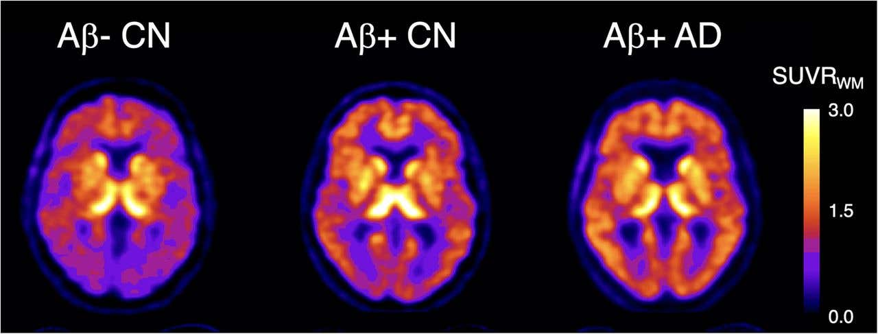
Graphical Abstract. 18F-SMBT-1 PET studies showed that Ab+ Alzheimer’s disease (AD) patients, but most importantly, Ab+ controls (CN) have significantly higher regional 18F-SMBT-1 binding than Ab- CN, with 18F-SMBT-1 retention highly associated with Ab burden. These findings suggest that increased 18F-SMBT-1 binding is detectable at the preclinical stages of Ab accumulation. (CREDIT: The Journal of Nuclear Medicine)
A new highly selective PET imaging agent can detect the presence of overexpressed monoamine oxidase-B (MAO-B) in cognitively unimpaired individuals with high beta amyloid (Ab)—one of the earliest signs of Alzheimer’s disease—according to research published in The Journal of Nuclear Medicine.
The radiotracer, 18F-SMBT-1, allows for a better understanding of the role of inflammation in Alzheimer’s disease, which can enable more accurate staging and prognosis at earlier stages.
Brain inflammation that accompanies Alzheimer’s disease involves reactive astrocytes, which are cells that overexpress MAO-B. The newly developed 18F-SMBT-1 radiotracer is highly selective for MAO-B and as a result has increased binding to reactive astrocytes.
“This increased binding suggests that 18F-SMBT-1 can potentially be used as a surrogate marker to detect reactive astrogliosis in Alzheimer’s disease,” noted Victor Villemagne, MD, professor of psychiatry at the University of Pittsburgh in Pittsburgh, Pennsylvania.
Related Stories
The study aimed to characterize 18F-SMBT-1 binding to reactive astrocytes across the Alzheimer’s disease continuum. Study participants included three clinical groups: 57 cognitively unimpaired controls, 12 subjects meeting criteria for mild cognitive impairment (MCI), and eight subjects meeting criteria for Alzheimer’s disease.
Participants underwent several types of imaging, including 18F-SMBT-1 PET, Ab PET, tau PET, and MRI. Images were normalized and statistical analyses conducted to assess 18F-SMBT-1 binding in relation to Ab and tau pathology burden. 18F-SMBT-1 was found to be highly correlated with Ab burden, and much less with tau burden.
The three clinical groups were then classified based on their Ab status (either as Ab+ or Ab-). No significant differences in 18F-SMBT-1 binding were found among Ab- participants in the control and MCI groups. In the Ab+ subjects with Alzheimer’s disease, 18F-SMBT-1 binding was significantly higher. Most importantly, 18F-SMBT-1 binding was significantly higher in the Ab+ control group as compared to Ab- control group.
Graphical Abstract. 18F-SMBT-1 PET studies showed that Ab+ Alzheimer’s disease (AD) patients, but most importantly, Ab+ controls (CN) have significantly higher regional 18F-SMBT-1 binding than Ab- CN, with 18F-SMBT-1 retention highly associated with Ab burden. These findings suggest that increased 18F-SMBT-1 binding is detectable at the preclinical stages of Ab accumulation. (CREDIT: The Journal of Nuclear Medicine)
“It’s of note that the brain regions where we saw this higher 18F-SMBT-1 binding in the control group are regions known for early Ab deposition. This suggests that reactive astrocytes are associated with early Ab deposition at the preclinical stages of Alzheimer’s disease and likely play a role over clinical progression,” said Villemagne.
He continued, “Implementation of 18F-SMBT-1 will clarify the role of reactive astrogliosis in neurodegenerative conditions, not just Alzheimer’s disease and its potential independent and/or synergistic effects on pathology, neurodegeneration, cognition, and disease progression. This has the potential to define and refine the diagnostic, staging and prognostic roles of reactive astrogliosis in these conditions.”
How does PET imaging work?
A positron emission tomography (PET) scan is an imaging test that can help reveal the metabolic or biochemical function of your tissues and organs. The PET scan uses a radioactive drug (tracer) to show both normal and abnormal metabolic activity.
Positron emission tomography: During a positron emission tomography (PET) scan, you lie on a narrow table that slides into a doughnut-shaped hole. The scanner takes about 30 minutes to produce detailed images of metabolic activity in your tissues and organs. (CREDIT: Creative Commons)
A PET scan can often detect the abnormal metabolism of the tracer in diseases before the disease shows up on other imaging tests, such as computerized tomography (CT) and magnetic resonance imaging (MRI).
The tracer is most often injected into a vein within your hand or arm. The tracer will then collect into areas of your body that have higher levels of metabolic or biochemical activity, which often pinpoints the location of the disease. The PET images are typically combined with CT or MRI and are called PET-CT or PET-MRI scans.
Note: Materials provided above by Society of Nuclear Medicine and Molecular Imaging. Content may be edited for style and length.
Like these kind of feel good stories? Get the Brighter Side of News' newsletter.