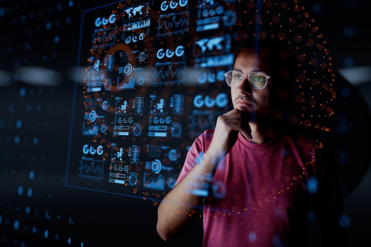
Researchers develop new tools for early cancer detection and treatment
New tissue-engineered models allow researchers to watch the earliest stages of cancer and may help doctors diagnose the disease much sooner.

 Edited By: Joseph Shavit
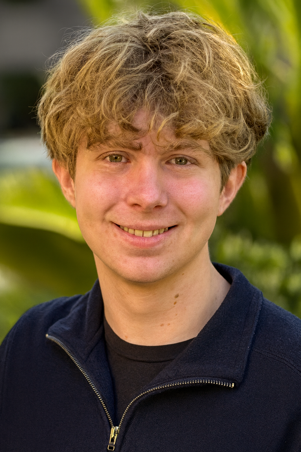
Haylie Helms, M.S., an OHSU graduate student in biomedical engineering, is lead author on a review in Nature Reviews. Helms works in the lab of Luiz Bertassoni, D.D.S., Ph.D., studying single-cell 3D bioprinting as a tool for early cancer detection. (CREDIT: OHSU/Christine Torres Hicks)
Cancer often infiltrates a person’s life long before anyone knows it. By the time symptoms arise and an examination indicates the worst, the disease has often been growing for months and sometimes years.
Many people refer to those early moments after an individual is diagnosed with cancer as stepping into a new and strange land. Questions come flooding in. How did this happen? Would it have been found sooner? Scientists have been asking those very questions for decades, yet the earliest moments of cancer remain obscured.
An important new scientific review is now shining a light on how engineers and biologists are working together to study cancer at its starting point. In a review published in the journal Nature Reviews Bioengineering, the authors highlight the wave of laboratory tools recently used to recreate the early stages of cancer with more accuracy than older laboratory approaches. These lab-based technologies use human cells rather than animal tissue and may ultimately give clinicians the ability to visualize more clearly how healthy tissue looks as it transforms into a dangerous disease.
Bridging the Gap Left by Missing Early Samples
For years, researchers have had little approach to tissue from very early tumors. Those first abnormal cell clusters are small and hard to find. When they are noted, they are often removed as quickly as possible and are rarely analyzed further. With organs deep within the body, accessing early tissue samples is frequently not even an option.
Without being able to witness the earliest biological changes, researchers are left with few options. Luiz Bertassoni, D.D.S., Ph.D., the senior author of the review and director of the Knight Cancer Precision Biofabrication Hub at Oregon Health & Science University, stated that accessibility to early-stage tissue has limited the field's progress.
"Early detection is one of the most critical variables in surviving cancer,” he stated. "These new technologies allow us to observe how cancer forms and process from the very beginning, making early cancer truly understandable; creating the potential for early diagnosis and even prediction of cancer initiation."
Engineering Miniature Models to Observe Cancer
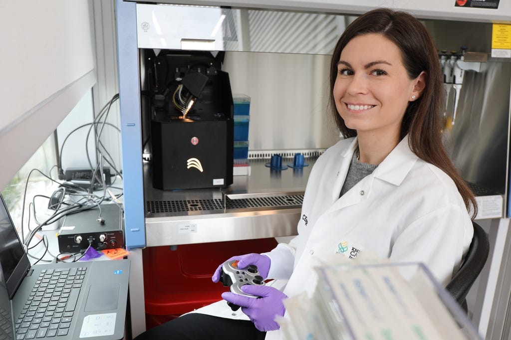
The review article highlights how engineers have been able to use 3D bioprinting, organoids, and organ-on-chip systems to model the complex environments present in human organs. These systems create tiny, realistic, and biologically active tissues that scientists can watch cancer emerge from, in real time.
3D bioprinting is exceptionally powerful. It utilizes known techniques from 3D printing to add cells in precise spatial patterns. This allows for the ability to grow tissues that recapitulate more of the biological behavior of tissues in the body.
The lead author Haylie Helms, M.S., a graduate student in biomedical engineering at OHSU, is studying how single-cell 3D bioprinting can be leveraged to understand and treat early disease. Helms is working on approaches to grow tissues that are healthy and then perturb that tissue toward precancerous or cancerous status.
"We can first build a healthy tissue and use different tools to turn it into cancer. "We can also take live cancer cells from the patient's biopsy and add these to the model," Helms said. "We can watch and say, ‘Why does a precancerous lesion in one person stay precancerous, and in another person, it becomes a malignant tumor?’"
Organoids—small clusters of cells developed from stem cells—recapitulate important characteristics of human organs. Organs-on-chips mimic blood flow, nutrient delivery, and mechanical forces such as pressure and stretch. Engineers can even manipulate the stiffness of the surrounding tissue, which is key because many cancers develop in locations that stiffen with time.
When immune cells are included, these systems start to represent the complete biological theatre of early cancer: competing for nutrients, immune responses, and subtle cues, directing some lesions towards aggression.
A New Way of Observing the First Signs of Disease
One goal shared among multiple labs is to use healthy cells and allow them to progress naturally through the earliest stages of cancer. They can change one variable at a time—such as chemical signals, mechanical stress, or genetic triggers—allowing them to target what pushes cells off their typical trajectory.
Some teams are now able to control precisely when genes related to neoplasia turn on. Light activation and ultrasound-based means permit scientists to turn those genes on in a precisely defined location and observe the first aberrant cells emerge. These models are repeatable, facilitating confirmation of findings from lab to lab.
This change demonstrates a significant increase in New Approach Methodologies, involving human-relevant systems to decrease dependence on animal testing. Many, if not most, of the tools were developed at the OHSU Knight Cancer Institute, where Bertassoni's earlier work on 3D printing blood vessels laid the groundwork.
With Respect to Earlier Detection and Cancer Interception
These platforms do more than show cancer inception. They also help scientists look for biomarkers, the subtle biological indicators that a tumor is forming. Validating an early biomarker would allow practitioners to identify cancer long before symptoms.
Some engineered systems can be personalized. By adding cells from a patient's biopsy, scientists can develop a model of that person’s biology. This would permit studies on how cells behave, model therapeutic development, and explore ways to prevent disease.
This notion is referred to as interception and is gaining momentum. Helms noted that while her vision is a worthy endeavor, most work is still around advanced disease. "A lot of the field is about late-stage cancer," she stated. "We want to understand and treat at the earliest moment possible."
Research findings are available online in the journal Nature Reviews Bioengineering.
Related Stories
- Early and accurate cancer detection now possible with $4 paper-based device
- Groundbreaking AI technology could improve early breast cancer detection by 30%
- Cutting-edge AI is transforming deadly skin cancer detection
Like these kind of feel good stories? Get The Brighter Side of News' newsletter.
Joshua Shavit
Writer and Editor
Joshua Shavit is a NorCal-based science and technology writer with a passion for exploring the breakthroughs shaping the future. As a co-founder of The Brighter Side of News, he focuses on positive and transformative advancements in technology, physics, engineering, robotics, and astronomy. Joshua's work highlights the innovators behind the ideas, bringing readers closer to the people driving progress.