Revolutionary brain imaging system to reveal how memories are made and recorded
Every day the brain makes and recalls new memories, yet we are still in the early stages of understanding this phenomenal process.
[July 11, 2023: Staff Writer, The Brighter Side of News]
Every day the brain makes and recalls new memories, yet we are still in the early stages of understanding this phenomenal process. (CREDIT: Creative Commons)
In the world of neuroscience, the complex dance of memory formation and recall remains a mystery that scientists relentlessly seek to unravel. With technology that has traditionally allowed only a limited view into the brain's intricate processes, the brain's capacity to generate and revisit memories is still not wholly understood.
However, an innovative imaging system developed by researchers at Michigan State University (MSU) offers an unprecedented window into the brain, promising to revolutionize our understanding of memory and cognition.
"Every day the brain makes and recalls new memories, yet we are still in the early stages of understanding this phenomenal process," explains Mark Reimers, an associate professor in the College of Natural Science and Institute for Quantitative Health Sciences and Engineering at MSU. "Our latest innovation will equip us with high-resolution images across the brain, providing an unparalleled level of detail and transforming our approach to neuroscience."
Delving into the intricacies of memory creation and retrieval is a fundamental quest for neuroscience. For Reimers, this pursuit also encompasses a desire to comprehend the issues faced by those suffering from memory disorders. He aims to elucidate the intricate processes underlying memory formation, its deterioration in conditions such as Alzheimer's disease, and the mishaps that occur in everyday memory.
Related Stories
"We want to know how memories are made and how they fail to be made in people with memory disorders like Alzheimer’s disease," says Reimers, highlighting the mission of his team. "We are especially intrigued by the opportunity to investigate and track the evolution of a memory over time and even observe how things get mixed up in everyday memory.”
Current brain imaging technology, despite being sophisticated, has limitations, especially in terms of resolution. The techniques used today can capture only a few hundred individual neurons — the nerve cells that transmit electrical signals throughout the body — at a time. Reimers, together with his co-investigator Christian Burgess from the University of Michigan, was determined to break these boundaries.
With seed funding from the director of IQHSE, Christopher Contag, and MSU’s neuroscience program, Reimers and Burgess set out to develop a ground-breaking imaging system. Their ambitious project aimed to image an astounding 10,000 to 20,000 neurons at once, offering an unparalleled view of brain activity in real-time as it forges and retrieves memories.
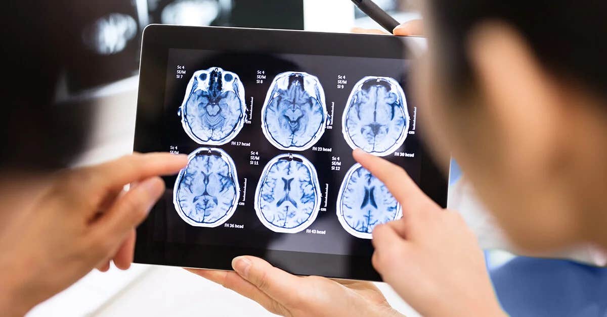
Brain image scan. (CREDIT: MSU)
The innovative endeavor caught the attention of the Air Force Office of Scientific Research, which awarded them a three-year $750,000 grant to support their groundbreaking research.
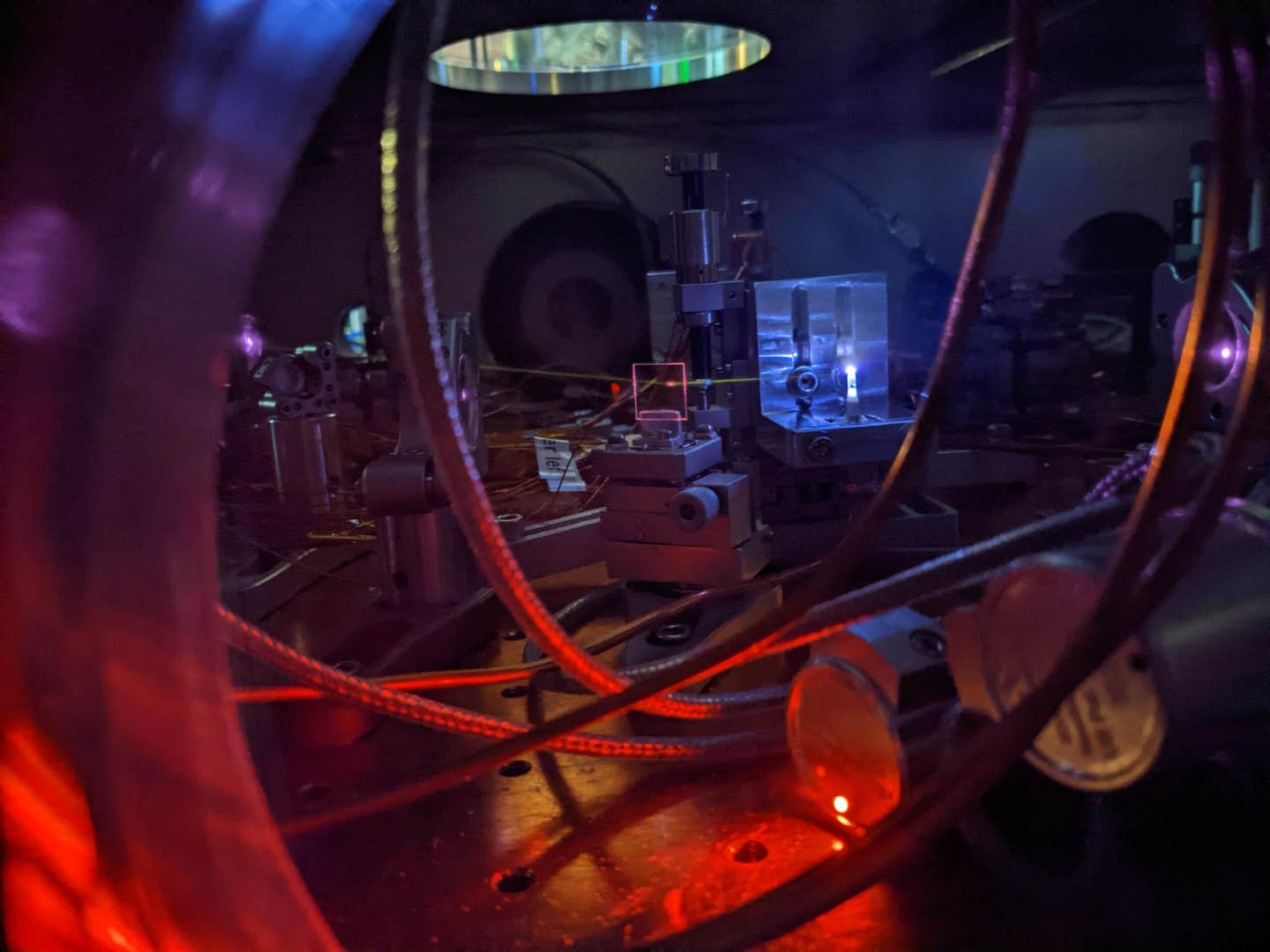
The newly-developed imaging system introduces a unique approach, employing a specially designed lens attached to a microscope. This microscope oscillates vertically between different planes, snapping multiple images every second. The resulting pictures show neurons firing in the outer layers of the brain's cortex, capturing the brain's activity during memory creation and recall.
In order to stimulate memory formation during their research, mice are exposed to specific series of sights, smells, and sounds. "A long-term goal of neuroscience has been to record from a great number of neurons as an animal is doing an activity and try to understand the relationship between the specific neurons that are firing at one point and what the animals are doing," explains Reimers. "We know that specific neurons are active at specific times, and those are correlated with what the animals are doing or what the animals are experiencing.”
A promising aspect of this cutting-edge technology is its potential to be paired with advanced image processing software. The research team aims to identify specific neurons employed by animals to create and recall memories. As Reimers anticipates, “We hope we will be the first people to observe and document memory formation across multiple regions of the cortex.”
The potential of this revolutionary
technology is not just confined to MSU. Reimers and Burgess hope that their system will prove invaluable to researchers worldwide, enabling them to improve the image quality for their projects and thereby advance the field of neuroscience.
“It cost us about $50,000 to build and we’re hoping that hundreds of different labs who are not particularly well funded will be able to use this system to do really cutting-edge neuroscience research,” Reimers shares. “Also, I would be happy to talk with other MSU labs who may want to use our new technology for their own research.”
The new imaging system developed by the researchers at MSU signifies a giant leap forward in neuroscience. It provides an exceptional window into the brain's labyrinthine processes of memory formation and recall, thereby contributing to our understanding of both normal brain function and disorders like Alzheimer’s disease. This innovative technology offers a fresh perspective on the human brain, unraveling its complexities and mysteries, and illuminating the science of memory like never before.
For more science stories check out our New Discoveries section at The Brighter Side of News.
Note: Materials provided above by The Brighter Side of News. Content may be edited for style and length.
Like these kind of feel good stories? Get the Brighter Side of News' newsletter.