Scientists create color X-ray images of the future
Sandia scientists unveil colorized X-ray imaging that sharpens detail and boosts medical, industrial, and security applications.
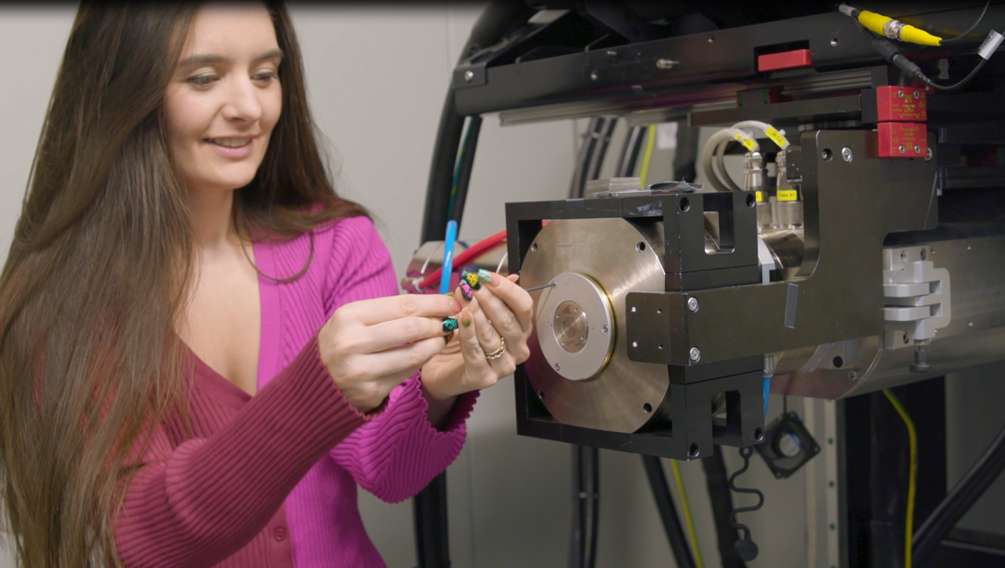
Noelle Collins mounts the packaged multi-metal patterned target on the X-ray tube at Sandia National Laboratories. (CREDIT: Mark Means)
When Wilhelm Röntgen found X-rays in the late 1800s, the world was given a new way of seeing inside the human body and peering into hidden structures. That black-and-white imaging method still powers everything from broken bone scans to airport checkpoints today.
But a group of scientists at Sandia National Laboratories believes they've found a way to add color, depth and accuracy to what has traditionally been a two-dimensional sight.
Project head Edward Jimenez is joined by materials scientist Noelle Collins and electronics engineer Courtney Sovinec. Together, they're building what they call "colorized hyperspectral X-ray imaging with multi-metal targets," or CHXI MMT. The name is technical sounding, but the concept is easy to grasp—adjust X-rays so they are more defined and colorful and therefore display more information.
From Black and White to Color
Traditional X-rays are created when a high-voltage electron strikes a solitary metal fragment, or an anode. Doing so, it releases X-rays that pass through an object and create a shadowy image detected by a sensor. Dense material like bone will absorb more rays, while softer tissue lets more pass through and creates the classic black-and-white image.
The problem, according to Collins, is that the images have limits. "With this new technology, we are essentially moving from the ancient way, which is black and white, to an entirely new colored world where we can more readily identify materials and defects of interest," she said.
The group came at the problem by minimizing the beam's focal spot. A reduced spot creates a sharper image, similar to a camera lens. To achieve that, they speckled the anode with dots of metal tungsten, molybdenum, gold, samarium and silver.
How Metals Produce Color
Each of those metals emits a unique "color" of X-ray light when electrons strike them. By positioning the dots so that they're smaller than the incoming beam, the group not only minimized the focal point but created a spectrum of X-ray colors.
Sovinec explained what follows: "Each metal generates a unique 'color' of X-ray light. When combined with an energy discriminating detector, we can detect individual photons, which provide us with density information and the energy of the photons. This allows us to identify the composition of the sample."
The outcome is an X-ray image with vivid clarity and far greater detail regarding the object itself. Jimenez summarized it: "We get a better representation of the shape and definition of that object, which is going to allow us to make unprecedented measurements and unprecedented observations."
Beyond Clearer Pictures
Good photographs are worth much more outside a laboratory. The researchers consider this to be a widely used technology. Security personnel may detect dangerous materials faster, factories may detect defects in machinery before they cause failures, and engineers may qualify parts by analyzing them without needing to take them apart.
The greatest assistance can potentially be offered to the medical field. By revealing slight changes in tissue density, doctors could identify medical issues much earlier. "You can see even minor differences with this technology," Jimenez explained. "We hope this will be used to spot things like cancer and more closely examine tumor cells."
In mammography, he explained, it can be extremely difficult to spot microcalcifications—minute points that could signal early breast cancer. Existing images tend to blur them or even overlook them. Colorized X-rays, however, render those subtle features more distinct and may improve early detection.
Looking Ahead
The Sandia researchers warn that their work is still just beginning, but the promise is clear. They plan to refine their design, test it in different industries, and collaborate with medical professionals, manufacturing and security experts. Collins summed it up in a few words: "From here we will continue to innovate. We hope to identify threats faster, diagnose diseases quicker and hopefully create a safer, healthier world."
This research can change your perspective on everything from airport security checks to body scans. For physicians, it will identify diseases sooner and more accurately, including cancers that are hard to find in their earliest stages.
For manufacturing and other sectors, clearer X-rays could make them safer by detecting flaws in materials before accidents occur.
Security experts can use it to detect threats more regularly. In short, the technology offers a pathway towards healthier lives, safer working places and stronger protections.
Related Stories
- Scientists temporarily turn skin transparent - eliminating the need for X-rays and CT scans
- New X-ray scans track violent solar winds threatening Earth’s satellites
- Scientists’ report world’s first X-ray of a single atom
Like these kind of feel good stories? Get The Brighter Side of News' newsletter.
Joseph Shavit
Writer, Editor-At-Large and Publisher
Joseph Shavit, based in Los Angeles, is a seasoned science journalist, editor and co-founder of The Brighter Side of News, where he transforms complex discoveries into clear, engaging stories for general readers. With vast experience at major media groups like Times Mirror and Tribune, he writes with both authority and curiosity. His writing focuses on space science, planetary science, quantum mechanics, geology. Known for linking breakthroughs to real-world markets, he highlights how research transitions into products and industries that shape daily life.