Scientists discover critical cell behavior driving cancer, immunity, and healing
Researchers found internal cell currents that rapidly push proteins to the leading edge, changing how cell movement is understood.

 Edited By: Joseph Shavit
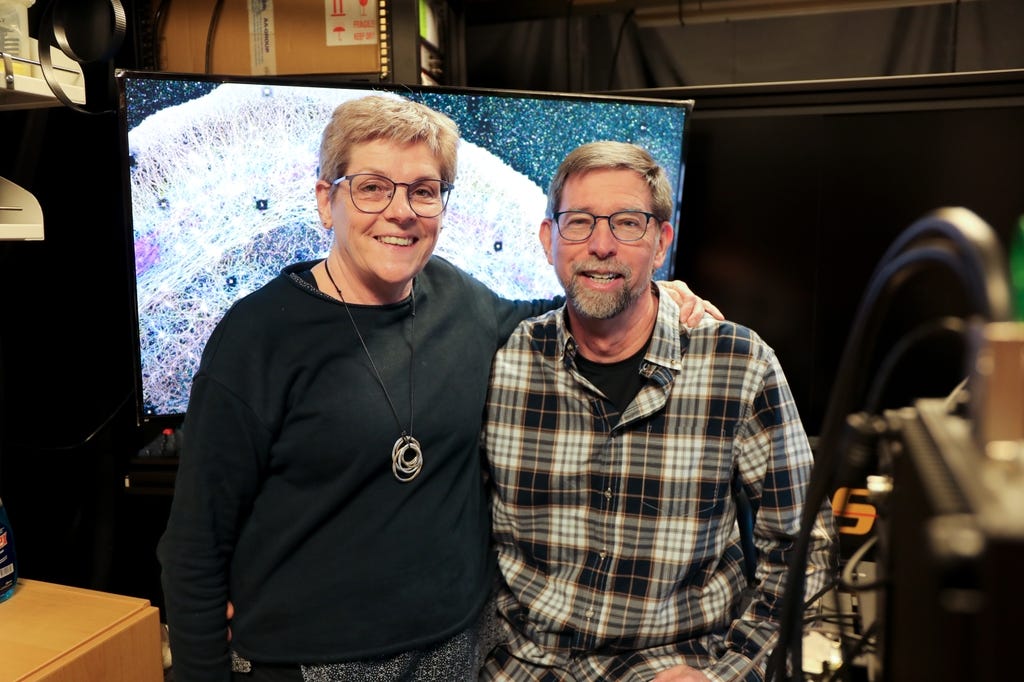
Cathy Galbraith, Ph.D., and Jim Galbraith, Ph.D., have a new study showing for the first time that cells create targeted streams of fluid to push proteins where they need to go. (CREDIT: OHSU/Christine Torres Hicks)
A moving cell looks simple from a distance. One edge pushes forward, the rest follows, and the whole thing creeps along as if its contents somehow know where to go.
That neat picture leaves out a crucial detail, according to a new study from Oregon Health & Science University. Inside the cell, proteins are not just drifting randomly until they reach the right spot. Instead, the researchers found what they describe as internal "trade winds." These are streams of fluid that push key proteins toward the cell’s leading edge. This part drives movement, repair and attachment.
The work, published in Nature Communications, challenges a long-standing assumption in biology. For years, textbook diagrams have treated many free-floating proteins as passengers of diffusion, moving by chance through the cell’s interior. This study argues that, at least in migrating cells, chance is not doing all the work.
The clue came from a dark line
The finding began with an accident. Catherine Galbraith and James Galbraith, both associate professors in OHSU’s Biomedical Engineering Department and Discovery Engine Investigators in the OHSU Knight Cancer Institute, were teaching a neurobiology course at the Marine Biological Laboratory in Massachusetts when something odd happened during a standard experiment.
“It actually started out as an unexpected finding,” Cathy Galbraith said. “We were just conducting an experiment with students in class.”
The team had used a laser to bleach proteins in a strip across the back of a living cell, making them temporarily invisible. Then a second thin dark line appeared near the front edge of the cell. That was not supposed to happen.
“We kind of did it for fun and then realized this gave us a way of measuring something that wasn’t able to be measured before,” she said.
That line turned out to mark soluble actin moving rapidly to the cell front. Actin is one of the main proteins that helps cells change shape and crawl. The researchers measured an apparent forward velocity of 3.6 ± 1.1 micrometers per second. This is nearly 50 times faster than the rearward movement of the original bleach line, which was measured at 0.08 ± 0.02 micrometers per second.
More than random drift
To probe what was driving that motion, the team used several imaging methods, including photobleaching, localized photoactivation, single-molecule tracking, fluorescence correlation spectroscopy, 3D structured illumination microscopy and iPALM, a super-resolution imaging technique.
They also built an inverse version of a standard fluorescence test and gave it a playful name: FLOP, for Fluorescence Leaving the Original Point.
“It wasn’t a flop at all,” Cathy Galbraith said. “Rather, it was the opposite. It is anything but a flop, because it worked.”
Using these tools in NG108 cells, CAD cells and 3T3 fibroblasts, the researchers found that myosin II contraction helps create a forward fluid flow. When they inhibited myosin activity, the directed movement weakened and became more symmetrical. This suggests that the cell’s internal machinery was no longer pushing proteins to the front as effectively.
“We realized the cartoon models in textbooks were missing a huge piece,” Jim Galbraith said. “There had to be some kind of flow in the cell pushing things forward. Cells really do ‘go with the flow.’”
The transport also appeared to be broadly non-specific. It did not move only actin. Proteins involved in protrusion and adhesion, including Arp3, vinculin and paxillin, also showed forward-biased movement toward the leading edge.
A front compartment with its own rules
The study goes further than a simple flow model. The researchers say the cell front acts like a distinct compartment, separated from the main cell body by an actin-myosin condensate barrier. It is not enclosed by a membrane. Therefore, they call it a pseudo-organelle.
That barrier seems to do two jobs. It slows exchange between the front and the rest of the cell, and it helps aim protein-rich flow toward the parts of the leading edge that are actively advancing.
“We found that the cell can actually squeeze at the back and target where it sends that material,” Jim Galbraith said. “If you squeeze half a sponge, the water only goes on that half. That’s basically what the cell is doing.”
The team says the curvature and position of these actin-myosin arcs can shift as the cell changes direction. In laser ablation experiments, disrupting a single arc caused the cell edge directly in front of it to collapse. This points to a local control system rather than a uniform one.
“Just as small shifts in the jet stream can change the weather, small changes in these cellular winds could change how diseases begin or progress,” Cathy Galbraith said.
The discovery could matter for cancer biology because invasive cells depend on rapid movement. Jim Galbraith said aggressive cancer cells may use this fast protein delivery system to feed protrusion at the front of the cell. “We know these highly invasive cells have this really cool mechanism to push proteins really fast, really rapidly where they need them at the front of the cell,” he said.
Still, the study was done in cultured cells using advanced microscopy, not in patients. The researchers say the work opens new directions for cancer research, wound healing, drug delivery and synthetic biology. But it does not by itself deliver a therapy.
“All you had to do was look,” Cathy Galbraith said. “The flows were there all along. Now we know how cells use them.”
Practical implications of the research
This work gives scientists a new way to think about how cells move and reorganize themselves so quickly.
By showing that cells can actively direct soluble proteins, rather than relying only on random diffusion, the study may help researchers look for weak points in migrating cancer cells.
It may also help them better understand tissue repair. Furthermore, the study might help design drug delivery or synthetic biology systems that borrow the same strategy.
Research findings are available online in the journal Nature Communications.
The original story "Scientists discover critical cell behavior driving cancer, immunity, and healing" is published in The Brighter Side of News.
Related Stories
- Scientists create advanced sense-and-respond systems that mimic natural cellular behavior
- Global first: AI tool predicts how proteins work in cells and tissues
- Researchers create new molecule to insert DNA into cells
Like these kind of feel good stories? Get The Brighter Side of News' newsletter.
Mac Oliveau
Writer
Mac Oliveau is a Los Angeles–based science and technology journalist for The Brighter Side of News, an online publication focused on uplifting, transformative stories from around the globe. Passionate about spotlighting groundbreaking discoveries and innovations, Mac covers a broad spectrum of topics including medical breakthroughs, health and green tech. With a talent for making complex science clear and compelling, they connect readers to the advancements shaping a brighter, more hopeful future.