Scientists found a previously unknown structure in mammalian cells
Scientists found a hidden organelle, the hemifusome, that may hold the key to how cells sort, recycle, and fight disease.
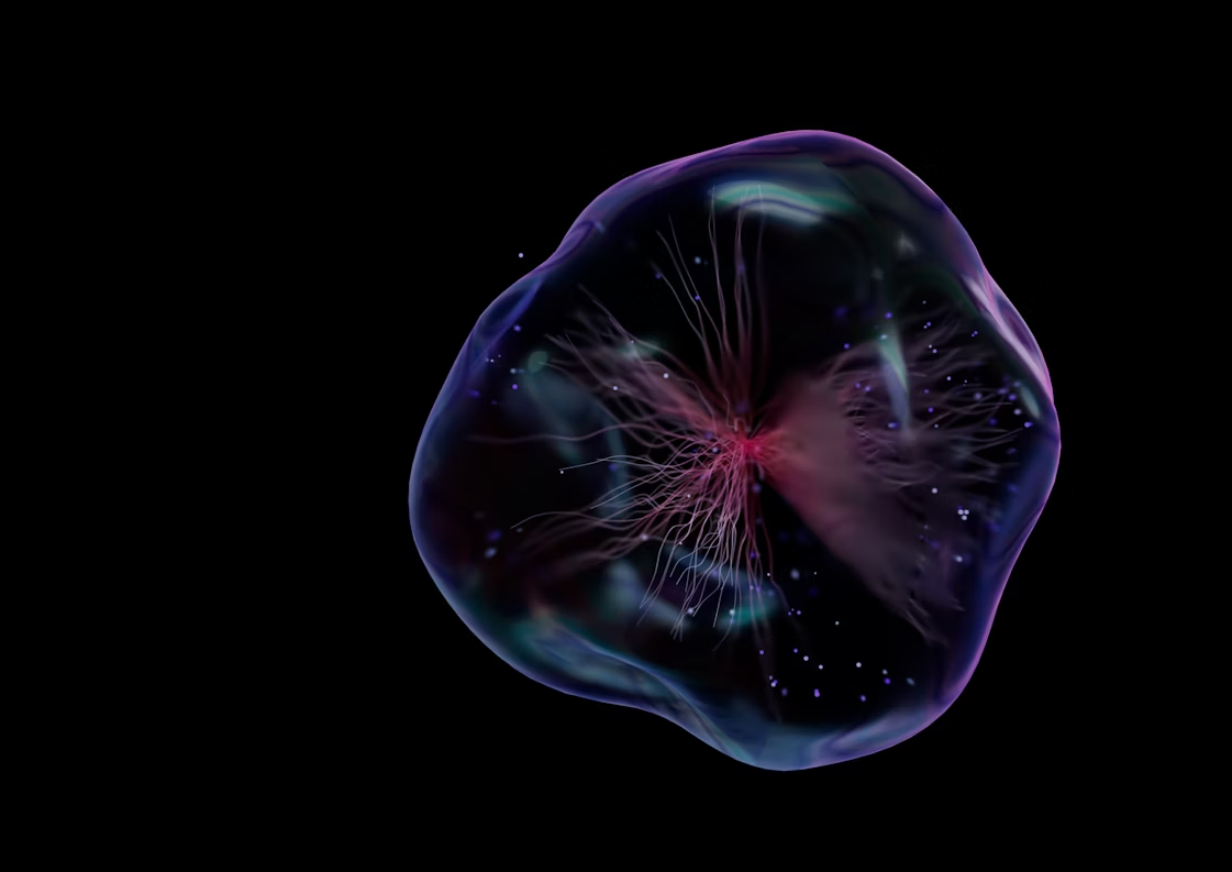
A new cell organelle called the hemifusome could help explain recycling problems behind complex genetic diseases. (CREDIT: Unsplash)
A new discovery inside your cells may change how scientists understand disease and design future treatments. Researchers have found a previously unknown structure in mammalian cells, something they believe plays a major role in keeping cells clean and organized.
They call this organelle the “hemifusome.” It helps your body’s cells sort, package, recycle, and get rid of materials they no longer need. Though small, this new organelle may hold huge importance in how our bodies work—and how they break down in disease.
A Tiny Organ That Does a Big Job
Scientists from the University of Virginia used cryo-electron tomography, or cryo-ET, to find the hemifusome. This advanced tool takes detailed images of cells in a near-natural state by freezing them. Unlike older methods, cryo-ET captures fast and delicate events inside cells without damaging them.
The scientists focused on a group of vesicles—small, sac-like structures inside cells that transport and organize materials. During the study, they discovered a new kind of vesicle structure that looked and acted differently from others.
These strange vesicles were stuck together in a way scientists had never seen before. They noticed that instead of fully fusing or staying apart, some vesicles were in a state called “hemifusion.” This means the outer layers of their membranes were joined, but the inner layers stayed separate.
They found these unusual vesicles shared a flat surface called a “hemifusion diaphragm” and were always attached to a 42-nanometer blob made of proteins and fats. The researchers named this structure a proteolipid nanodroplet, or PND. Together, these parts form what the scientists have named the hemifusome.
Up to 10% of all vesicular organelles at the edges of cells belong to this new structure. The hemifusome doesn't take part in normal endocytosis—the usual way cells absorb materials. Instead, it follows its own path, forming new vesicles and even multi-vesicle structures.
Related Stories
- Breakthrough new technology can freeze time for cells
- New bio-engineered molecule kills cancer cells and activates the immune system
Uncovering a New Path in Cell Recycling
The team believes the hemifusome helps create multivesicular bodies, or MVBs. These are special organelles that carry cargo meant for reuse or disposal. Normally, the cell uses a process called ESCRT to form MVBs. But the hemifusome may offer a different route—one that doesn’t depend on ESCRT. That could explain how some cells still manage to sort and recycle cargo even when ESCRT doesn’t work.
“This is like discovering a new recycling center inside the cell,” said researcher Seham Ebrahim, PhD, from the University of Virginia’s Department of Molecular Physiology and Biological Physics. “We think the hemifusome helps manage how cells package and process material, and when this goes wrong, it may contribute to diseases that affect many systems in the body.”
The shape and activity of the hemifusome vary from cell to cell. Some carry smaller vesicles inside them, showing they can change form and function. The researchers believe these structures act as flexible platforms for making and managing vesicles.
Ties to Inherited Disease
Problems with how cells move and recycle cargo can lead to disease. One such condition is Hermansky-Pudlak syndrome, a rare disorder that causes problems with skin color, vision, blood clotting, and lung function.
“We’re just beginning to understand how this new organelle fits into the bigger picture of cell health and disease,” Ebrahim said. “It’s exciting because finding something truly new inside cells is rare – and it gives us a whole new path to explore.”
The hope is that understanding the hemifusome will help scientists discover how certain diseases begin. For example, if the hemifusome doesn’t form properly, cells may fail to clean themselves up. Over time, this could lead to a buildup of waste that affects many parts of the body.
“You can think of vesicles like little delivery trucks inside the cell,” Ebrahim explained. “The hemifusome is like a loading dock where they connect and transfer cargo. It’s a step in the process we didn’t know existed.” Because the hemifusome helps shape how cells sort and dispose of material, it may become a key focus for treating diseases linked to poor waste management in cells.
The Science Behind the Images
The team worked across two major research groups: one at the University of Virginia, the other at the National Institutes of Health. Ebrahim, along with Dr. Bechara Kachar and scientists Amirrasoul Tavakoli and Shiqiong Hu, used cryo-ET to gather ultra-clear images of living cells.
In total, they studied four different types of mammalian cells. In all of them, they found the hemifusome appeared in different shapes but followed the same basic structure. They always saw the hemifused vesicles connected through the flat diaphragm and anchored by the PND. This repeated pattern showed the hemifusome was not just a glitch or rare event—it was real and common.
They also saw how the hemifusome avoided the usual endocytic path. It seemed to form and act independently. That’s important because it means cells may use more than one method to sort and send out cargo. If scientists can figure out how the hemifusome pathway works, they might be able to fix it when it breaks down.
Ebrahim and her team shared their findings in the respected science journal Nature Communications. Their detailed research lays the groundwork for what may become a whole new field in cell biology.
“This is just the beginning,” Ebrahim said. “Now that we know hemifusomes exist, we can start asking how they behave in healthy cells and what happens when things go wrong. That could lead us to new strategies for treating complex genetic diseases.”
By finding this new organelle, scientists have unlocked a new part of the puzzle. The hemifusome may help explain how cells stay clean, organized, and healthy—and what happens when that balance fails.
Note: The article above provided above by The Brighter Side of News.
Like these kind of feel good stories? Get The Brighter Side of News' newsletter.