World’s first living implant could help paralyzed organs move again
MIT’s new living implant rewires muscle to help restore movement in paralyzed organs.

 Edited By: Joseph Shavit
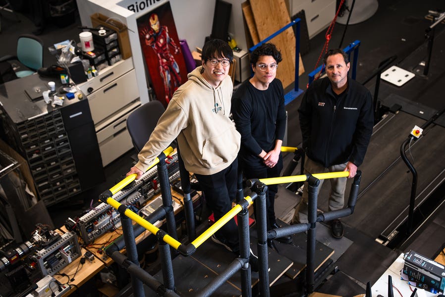
MIT researchers (from left to right) Hyungeun Song, Guillermo Herrera-Arcos, and Hugh Herr have developed the first “living” implant that uses rewired sensory nerves to revive paralyzed organs. (CREDIT: JimDay)
A muscle that no longer answers to the brain might sound useless. MIT researchers are trying to turn that idea into medicine.
In a study published in Nature Communications, the team described a new implant called a myoneural actuator, or MNA. It uses living skeletal muscle that has been surgically rewired so a computer, not the brain, can control it. In rodent tests, the system restored squeezing motion in the small intestine and also passed sensory signals back toward the brain, raising the possibility of future implants that could help revive organ function and restore some lost bodily sensations.
The work comes from researchers at the MIT Media Lab, the K. Lisa Yang Center for Bionics, and the McGovern Institute for Brain Research. Senior author Hugh Herr led the study with postdoctoral associate Guillermo Herrera-Arcos and former postdoc Hyungeun Song.
Rewiring muscle for a different job
The idea starts with a stubborn medical problem. Many organs rely on motion, but once the link between the nervous system and that motion breaks down, restoring it becomes very difficult. Mechanical devices can be bulky, hard to miniaturize, and poorly suited for the body. Lab-grown muscle, meanwhile, remains complex and far from clinical use.
So the MIT team took a different route. Instead of building a new actuator from scratch, they reworked existing muscle inside the body.
“We engineered existing muscles to become an actuator, or motor, that reinstates motion in organs,” Song said.
To do that, the researchers removed a muscle’s normal motor nerve input and replaced it with a sensory nerve. That sounds backward, but it served two purposes. First, it let the muscle remain living and functional. Second, it shifted command away from the brain and toward a computer-controlled system using functional electrical stimulation.
“You don’t want the brain to consciously control the muscle actuator because you want the actuator to automatically control an organ, like the heart,” Herrera-Arcos said.
A surprising nerve connection
One of the study’s central findings was that sensory nerves could do something scientists had not clearly established before in this context: reinnervate muscle and form working neuromuscular junctions.
“Remarkably, when the team replaced motor nerves in rodent muscle with sensory ones, the sensory nerves reinnervated the muscles and formed functional synapses. It’s a tremendous discovery,” Herrera-Arcos said.
The team used the lateral gastrocnemius muscle in rodents as the base actuator and the sural nerve for sensory reinnervation. Histology showed that the sensory nerve had smaller and more uniform axon sizes than the native motor nerve. That mattered because ordinary motor nerves tend to recruit large axons first under electrical stimulation, which tires muscles quickly.
Sensory nerves, by contrast, distribute the signal more evenly.
The payoff was striking. Under continuous stimulation, the rewired muscle showed a 260 percent improvement in fatigue resistance over native muscle, lasting 18.67 ± 2.96 seconds compared with 5.19 ± 1.16 seconds in the control group. In repeated stimulation tests, it also lost force more slowly and produced more stable output.
The researchers also found that once reinnervation was complete, the actuator’s mass and force stayed stable from 9 to 15 weeks after surgery without outside interventions, though some mass had been lost during the reinnervation process.
From amputations to the gut
The team then tested how this “living” actuator might work in biohybrid systems.
In one proof-of-concept setup, the researchers designed what they call a Proprioceptive Mechanoneural Interface, or PMI, for amputations. In rodents, they linked the rewired actuator to another muscle meant to mimic residual muscle in an amputated limb. As the actuator’s output increased, the strain in that end organ rose, and so did modulated neural afferent signals. That suggests the system may someday help prosthetic users receive more natural sensory feedback.
In another demonstration, the researchers wrapped the actuator around a rodent small intestine. Optical flow analysis showed that movement in the actuator and the intestine stayed strongly synchronized, indicating that the implant could mechanically modulate organ motion.
“This suggests that our technology could seamlessly link organs to the brain,” Song said. “For example, we might be able to make a paralyzed stomach relay hunger.”
The authors point to possible future uses in conditions that impair intestinal contractions, including ileus, diabetic enteropathy, and Crohn’s disease. They also mention longer-term possibilities involving the bladder, lungs, skin grafts, and even systems tied to virtual reality, where mechanical stimulation could be used to create forms of sensory feedback.
What still stands in the way
The study was done in rodents, and the researchers are clear that larger-animal testing and eventually human studies would be needed before any clinical use.
They also note open questions. The exact reinnervation mechanism still needs more study, including which nerve fibers are chiefly responsible for the cholinergic signaling seen in the actuator. The team says it is also necessary to evaluate long-term stability when the actuator is coupled to a target organ. In some cases, unintended sensations or pain from stimulation could require reversible nerve block. For applications involving the heart, electrical isolation would be critical because past work has shown dangerous rhythm problems can arise when skeletal and cardiac muscle interact improperly.
Still, the researchers argue that the approach may be more clinically practical than it first sounds. It relies on reconstructive surgical techniques already used in other settings, along with nerve cuff electrodes and pulse generators that are already common in medicine. Because it uses a patient’s own muscle, it may also avoid some of the infection, rejection, and immunosuppression issues tied to donor tissue or engineered implants.
Practical implications of the research
This study points to a different kind of medical device, one built from the patient’s own living tissue rather than a fully synthetic machine.
If the approach holds up in larger studies, it could offer a new way to help organs move again after nerve damage, improve sensory feedback for prosthetic users, and create biohybrid implants that work more naturally with the body.
Research findings are available online in the journal Nature Communications.
The original story "World's first living implant could help paralyzed organs move again" is published in The Brighter Side of News.
Related Stories
- Paralyzed man moves robotic arm with his mind
- Life-changing implantable tactile sensors offer new hope for paralyzed individuals
- Groundbreaking new spinal implant helps paralyzed people walk again
Like these kind of feel good stories? Get The Brighter Side of News' newsletter.
Mac Oliveau
Writer
Mac Oliveau is a Los Angeles–based science and technology journalist for The Brighter Side of News, an online publication focused on uplifting, transformative stories from around the globe. Passionate about spotlighting groundbreaking discoveries and innovations, Mac covers a broad spectrum of topics including medical breakthroughs, health and green tech. With a talent for making complex science clear and compelling, they connect readers to the advancements shaping a brighter, more hopeful future.