Flexible electronic device wraps around the spinal cord – revolutionizing the treatment of spinal injuries
A groundbreaking flexible electronic device that wraps around the spinal cord, potentially revolutionizes the treatment of spinal injuries
A team from the University of Cambridge has developed a groundbreaking flexible electronic device that wraps around the spinal cord, potentially revolutionizing the treatment of spinal injuries, which often result in severe disability and paralysis. This innovative device, created by engineers, neuroscientists, and surgeons, offers a comprehensive approach to recording and stimulating nerve signals between the brain and spinal cord, presenting a significant advancement over existing methods.
Unlike current treatments that require high-risk surgeries involving electrodes piercing the spinal cord or brain implants, the Cambridge-developed devices provide a safer alternative. These devices are capable of 360-degree recording, capturing a complete picture of spinal cord activity without invasive procedures. The findings were published in the journal Science Advances.
“The spinal cord is like a highway, carrying information in the form of nerve impulses to and from the brain,” explained Professor George Malliaras from the Department of Engineering, co-leader of the research. “Damage to the spinal cord causes that traffic to be interrupted, resulting in profound disability, including irreversible loss of sensory and motor functions.”
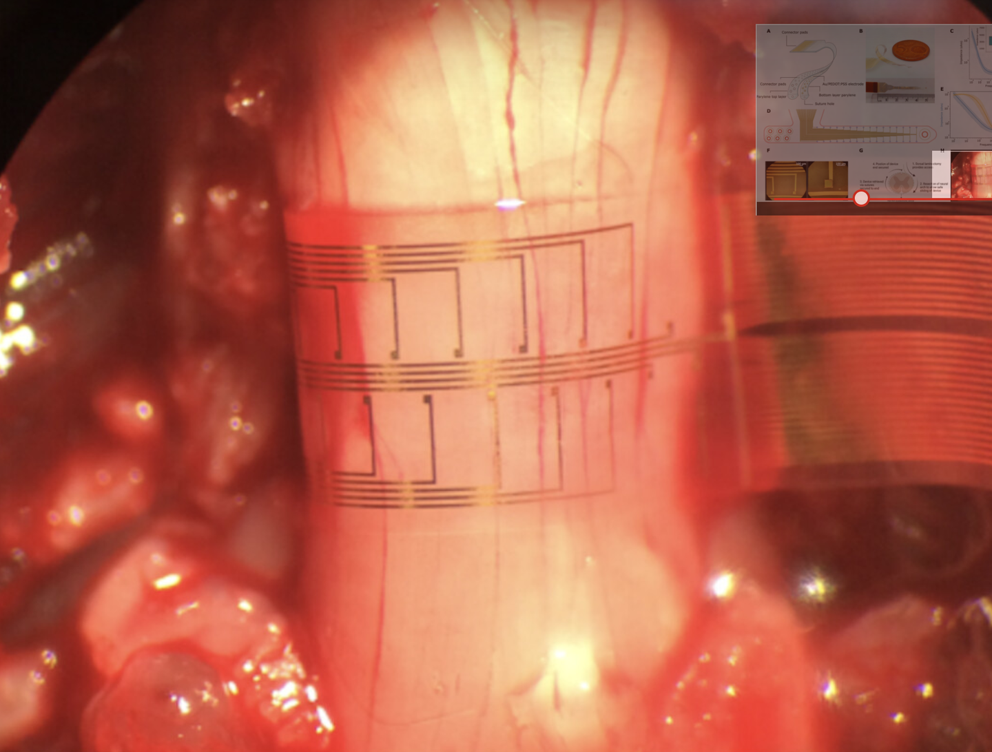
Dr. Damiano Barone from the Department of Clinical Neurosciences, also a co-leader, added, “Most technologies for monitoring or stimulating the spinal cord only interact with motor neurons along the back, or dorsal, part of the spinal cord. These approaches can only reach between 20 and 30 percent of the spine, so you’re getting an incomplete picture.”
The Cambridge team drew inspiration from microelectronics to create biocompatible devices, just a few millionths of a meter thick, using advanced photolithography and thin film deposition techniques.
These high-resolution implants wrap around the spinal cord, intercepting signals on the axons or nerve fibers, and recording them without causing damage due to their minimal thickness.
Related Stories:
“It was a difficult process because we haven’t made spinal implants in this way before, and it wasn’t clear that we could safely and successfully place them around the spine,” Malliaras noted. “But because of recent advances in both engineering and neurosurgery, the planets have aligned, and we’ve made major progress in this important area.”
In tests on live animal and human cadaver models, the devices demonstrated their ability to stimulate limb movement and bypass complete spinal cord injuries where communication between the brain and spinal cord had been entirely interrupted. These tests showed very low latency, meaning the devices' reaction time was close to human reflexive movement.
“If someone has a spinal injury, their brain is fine, but it’s the connection that’s been interrupted,” Barone explained. “As a surgeon, you want to go where the problem is, so adding brain surgery on top of spinal surgery just increases the risk to the patient. We can collect all the information we need from the spinal cord in a far less invasive way, so this would be a much safer approach for treating spinal injuries.”
While treatments using these devices are still several years away, the researchers believe they could soon be valuable for monitoring spinal cord activity during surgery. This could lead to improved treatments for a range of conditions, including chronic pain, inflammation, and hypertension. The ability to monitor spinal cord signals could also aid in developing better therapies for these conditions.
“It’s been almost impossible to study the whole of the spinal cord directly in a human, because it’s so delicate and complex,” said Barone. “Monitoring during surgery will help us to understand the spinal cord better without damaging it, which in turn will help us develop better therapies for conditions like chronic pain, hypertension, or inflammation. This approach shows enormous potential for helping patients.”
The researchers are planning to use the devices to monitor nerve activity in the spinal cord during surgery, providing insights into this complex and understudied part of human anatomy. Understanding the spinal cord better could pave the way for new treatments and therapies, offering hope for patients with various spinal conditions.
The research received support from several prestigious institutions, including the Royal College of Surgeons, the Academy of Medical Sciences, Health Education England, the National Institute for Health Research, and the Engineering and Physical Sciences Research Council (EPSRC), part of UK Research and Innovation (UKRI).
By offering a safer, less invasive method of recording and stimulating nerve signals, this new technology holds promise for transforming patient care and expanding our knowledge of spinal cord function and its associated conditions.
For more technology news stories check out our New Innovations section at The Brighter Side of News.
Note: Materials provided above by The Brighter Side of News. Content may be edited for style and length.
Like these kind of feel good stories? Get the Brighter Side of News' newsletter.