A ‘third eye’ helps fish navigate deep underwater
A brain region helps zebrafish combine light signals from eyes and a “third eye” to guide movement.

 Edited By: Joseph Shavit
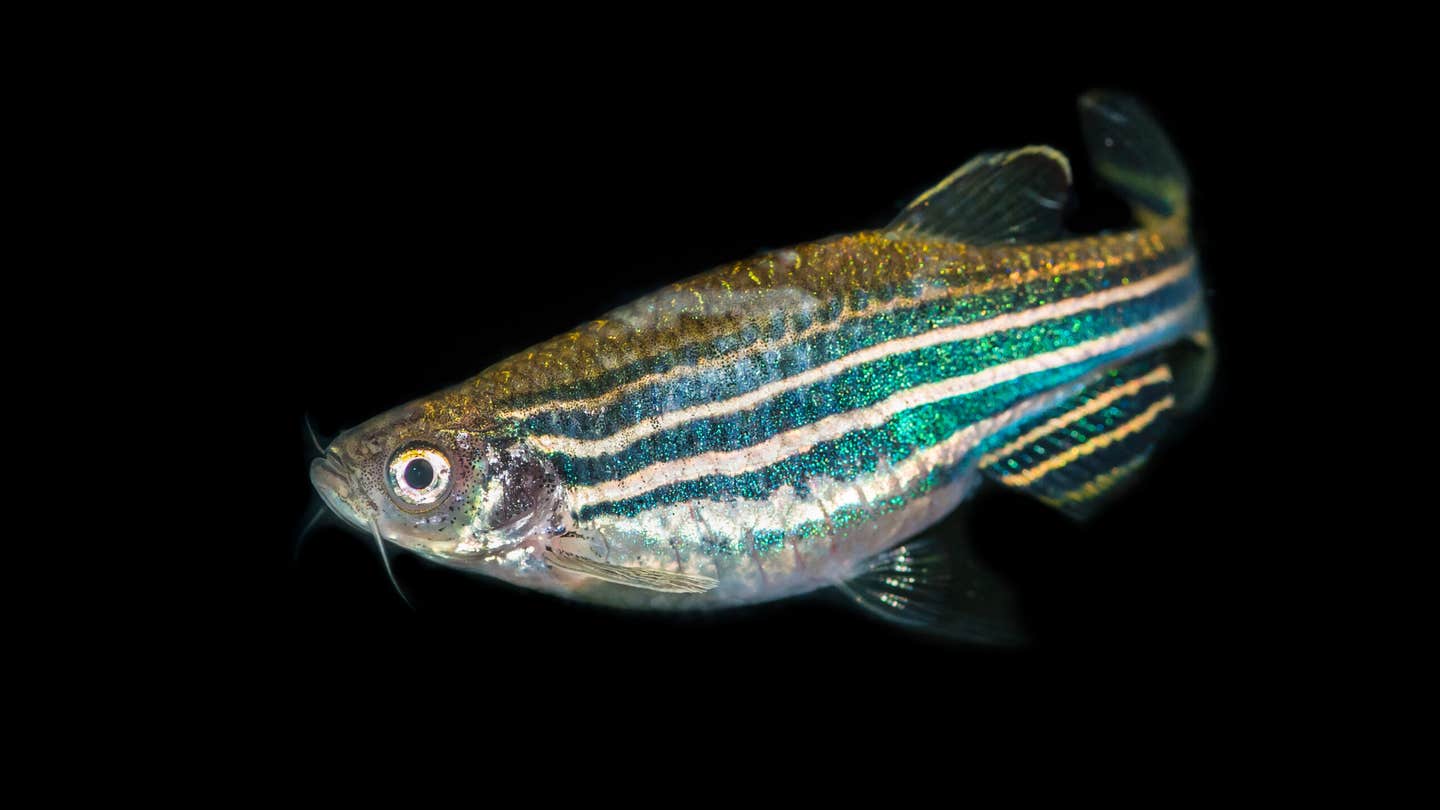
Zebrafish study reveals how brain integrates light signals to control vertical swimming behavior. (CREDIT: Shutterstock)
Light behaves differently underwater. It shifts with depth, bends through murky currents, and separates into distinct wavelengths that change from surface to seafloor. For fish, those subtle differences are not just background noise. They are cues.
A new study from Osaka Metropolitan University points to a specific region deep in the zebrafish brain that helps decode those cues. The work identifies the tegmentum, part of the midbrain, as a place where two streams of light information meet, one from the eyes and another from a lesser-known light sensor often called the “third eye.” Together, those signals appear to guide a simple but essential behavior: whether a fish swims up or down.
The finding adds detail to a long-standing question about how animals translate light into movement, especially in environments where light is constantly changing.
Where Two Light Systems Converge
Fish do not rely on a single visual pathway. Alongside their eyes, many species possess a pineal organ, a small structure sensitive to light that sits near the top of the brain. It detects environmental brightness and wavelength in ways that complement vision.
The research team, led by Professors Akihisa Terakita and Mitsumasa Koyanagi with Dr. Seiji Wada, focused on how these two systems communicate. Their attention centered on a protein called parapinopsin 1, or PP1, an opsin that responds to light. Opsins are common in eyes, but PP1 also operates in the pineal organ.
Instead of mapping the entire brain, the researchers traced how signals tied to PP1 travel. They followed activity from the pineal photoreceptor cells through nerve pathways and into the midbrain. That path led them to the tegmentum. There, signals from the eyes and the pineal organ come together. According to the study, this integration allows the fish to interpret differences in light wavelength, including contrasts between visible and ultraviolet light, and translate that information into vertical movement.
Watching Signals Move in Real Time
The team relied on a practical advantage of zebrafish larvae: transparency. Their bodies allow scientists to observe neural activity directly. “We decided to study zebrafish, as their larvae are transparent,” Professor Koyanagi said. “This transparency means that changes in calcium levels within nerve cells can be observed as changes in the fluorescence intensity of the calcium indicator, allowing us to measure the strength of neural activity.”
Using calcium imaging, the researchers tracked how neurons responded to light. When nerve cells become active, calcium levels shift, and those shifts can be visualized through changes in brightness. This method gave the team a way to watch signals move through the brain in response to different wavelengths.
PP1 played a central role in those responses. The protein reacts in opposite ways to ultraviolet and visible light. That contrast provided a clear signal for the researchers to follow. They traced these signals from the pineal organ, through ganglion cells, and into the tegmentum. Along the way, the signals retained their wavelength-specific information.
A Behavior Tied to Color
The integration of signals in the tegmentum does not stay abstract. It connects directly to behavior.
“Our study showed that the tegmentum integrates visual information from the eyes that is combined with color information detected by the pineal organ. These integrated signals then contribute to the fish’s up and down swimming behavior,” Dr. Wada said.
Fish live in an environment where light carries information about depth and conditions. Ultraviolet and visible light do not penetrate water in the same way. Differences in those wavelengths can signal whether a fish is near the surface or deeper below. By combining inputs from two light-detecting systems, the brain can form a more complete picture of those conditions. The result is a behavioral adjustment, swimming upward or downward in response to the light environment. It is a simple action, but it depends on a layered process.
What Happens When the Signal Breaks
To test how essential PP1 is, the researchers examined fish that lacked the gene responsible for producing it. Without PP1, the chain of signals from the pineal organ changes. Those fish did not respond to differences in light wavelength in the usual way. The typical vertical movements tied to light cues were absent.
This absence strengthens the idea that PP1-based signaling contributes directly to the behavior. It also reinforces the role of the tegmentum as a hub where these signals matter. Without the proper input, the system does not function as expected.
A System Built on Opposites
One of the more striking aspects of the study lies in how PP1 behaves. It does not respond to all light in the same direction. Instead, it produces opposite reactions depending on whether the light is ultraviolet or within the visible spectrum. That opposition creates contrast. It allows the nervous system to distinguish between types of light rather than simply measuring brightness.
The brain then combines that contrast with visual information from the eyes. The tegmentum becomes the place where these signals overlap, not as duplicates, but as complementary pieces of information. This layered input appears to give the fish a more nuanced sense of its surroundings.
A Closer Look at Neural Circuits
The study also touches on a broader goal in neuroscience: mapping how specific signals move through the brain and shape behavior.
“These findings shed light on how animals process visual information, advance the analysis of neural circuits using light, and expand research into behavioral control,” Professor Terakita said. “In the future, these findings may contribute to applications in neuroscience and biomedicine, such as the identification of neural circuits using PP1-based optogenetics.”
The mention of optogenetics points to a technique where light is used to control or trace neural activity. Proteins like PP1 could serve as tools in that work, helping scientists identify and manipulate specific circuits. While the current study focuses on zebrafish, the underlying approach has wider relevance. Understanding how light-sensitive proteins influence neural pathways could inform research beyond aquatic species.
A Small Brain Region with a Defined Role
The tegmentum does not dominate most discussions of the brain. Yet here it appears as a precise meeting point for two sensory streams. That specificity stands out. Instead of a diffuse network, the study highlights a defined region where integration occurs. Signals from the eyes and the pineal organ do not remain separate. They converge, combine, and influence movement.
The finding suggests that even relatively simple behaviors may depend on targeted processing areas rather than broad, generalized activity.
Practical Implications of the Research
This work clarifies how animals turn environmental light into action, using defined neural pathways and specialized proteins. By identifying where and how signals combine, the study offers a clearer map of sensory processing in the brain.
That map could support future efforts to study neural circuits in more detail, especially those linked to behavior. The use of PP1 and similar proteins may also expand tools for tracking or controlling brain activity with light.
Beyond fish, the findings contribute to a broader understanding of how multiple sensory systems integrate information. That principle applies across species, including in areas of neuroscience and biomedical research focused on perception and behavior.
Research findings are available online in the journal PNAS.
The original story "A 'third eye' helps fish navigate deep underwater" is published in The Brighter Side of News.
Related Stories
- Researchers develop plant-based cleanup for harmful antibiotics found in rivers and fish
- Fishing nets and recycled plastic trash are being paved into Hawaii's roads
- Astronomers spot 'jellyfish galaxy' torn apart 8.5 billion years ago
Like these kind of feel good stories? Get The Brighter Side of News' newsletter.
Hannah Shavit-Weiner
Medical & Health Writer
Hannah Shavit-Weiner is a Los Angeles–based medical and health journalist for The Brighter Side of News, an online publication focused on uplifting, transformative stories from around the globe. Passionate about spotlighting groundbreaking discoveries and innovations, Hannah covers a broad spectrum of topics—from medical breakthroughs and health information to animal science. With a talent for making complex science clear and compelling, she connects readers to the advancements shaping a brighter, more hopeful future.