Brain implant helps paralyzed man move his hand and feel touch again
Brain implants, spinal stimulation and AI helped a man with tetraplegia grasp objects, feed himself and regain limited touch.

 Edited By: Joseph Shavit

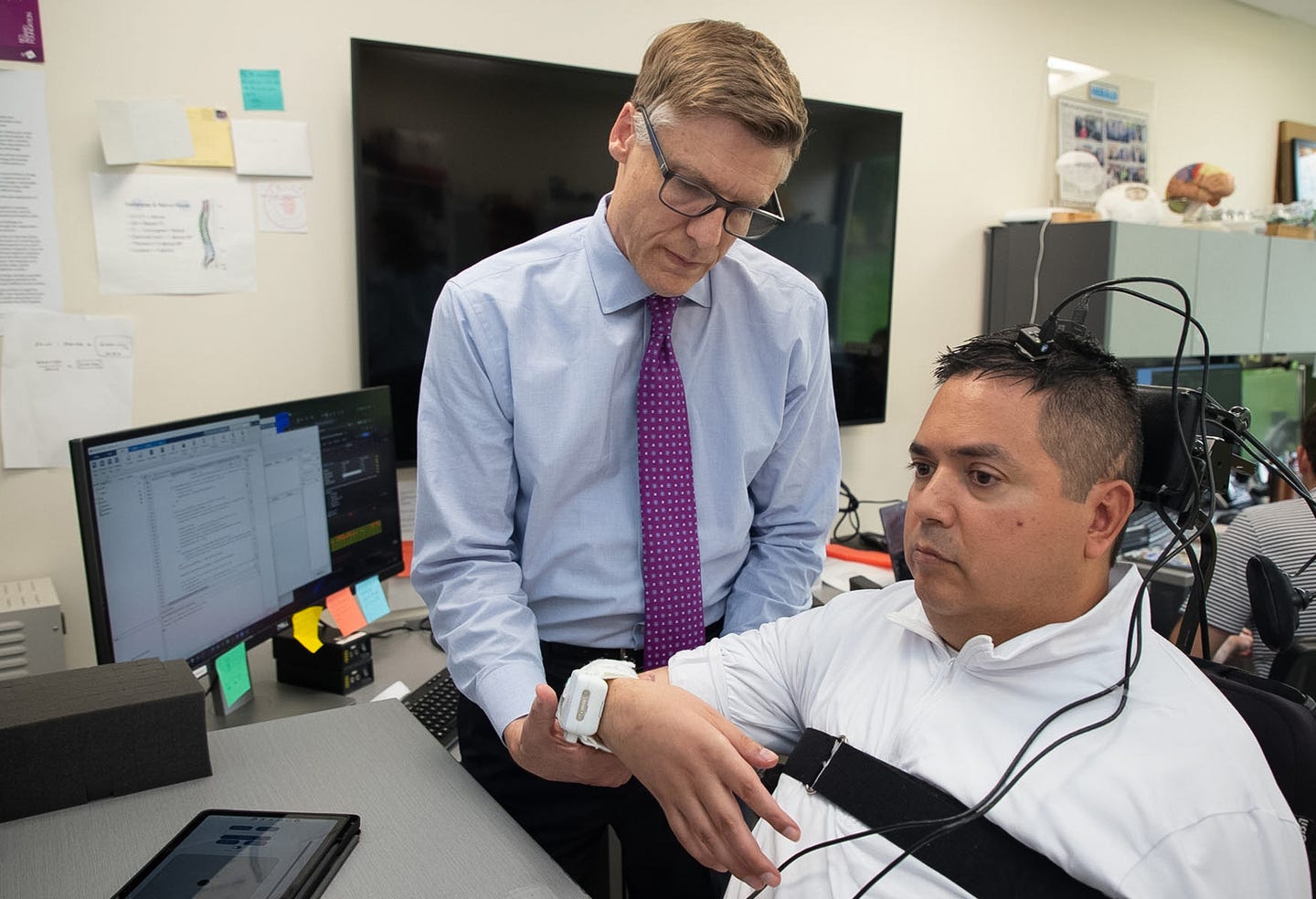
A neural bypass for paralysis restored hand movement, grip control and wrist sensation, with some benefits lasting after stimulation. (CREDIT: Feinstein Institutes for Medical Research)
A man paralyzed from the shoulders down can now bring his hands to his face, grasp a cup and feel pressure on his wrist. A brain and spinal cord system helped restore abilities once considered exceedingly difficult to recover.
The experimental “double neural bypass” combines brain implants, electrical stimulation, artificial intelligence and a wearable hand device. It reads the man’s intention to move, activates his own muscles and sends touch information back to his brain.
Some improvements continued when stimulation stopped. That result suggests the system could act as both an assistive device and a rehabilitation tool, according to research published in Nature Medicine.
The trial involved a 42-year-old man with complete tetraplegia following a diving accident. His injury occurred at the C4 sensory and C5 motor levels, leaving him unable to lift his hands to his face, grasp objects or feel his hands and lower forearms.
Building a bridge around the injury
Spinal cord injuries interrupt communication between the brain and the body. Complete injuries are especially difficult because voluntary movement and sensation are absent below the damaged area.
The system bypasses that interruption through several linked components. Two implanted microelectrode arrays record activity in the primary motor cortex, which controls voluntary movement. Three additional arrays stimulate the primary somatosensory cortex, the region that processes touch.
Algorithms translate recorded brain activity into commands for opening, closing or resting the participant’s right hand. Electrical patches on his forearm then stimulate muscles that flex and extend the fingers.
A custom spinal patch delivers noninvasive stimulation through the skin over selected cervical spinal roots. The researchers paired this stimulation with repeated attempts to move, aiming to strengthen remaining communication pathways.
Spinal stimulation alone produced meaningful gains in the participant’s arms. After 15 weeks, voluntary elbow-flexion force increased by 61 percent on the right and 25 percent on the left.
After about 35 weeks, the gains reached 86 percent on the right and 62 percent on the left. The improvements allowed him to lift both hands to his face, an action he could not perform before treatment.
Brain signals reopen the hand
Spinal stimulation did not restore voluntary finger movement or improve feeling in the hands. The team therefore activated the system’s brain-controlled assistive components.
The participant watched a virtual hand while attempting to open, close or rest his own hand. A long short-term memory neural network learned to recognize the corresponding patterns in his motor cortex.
Only signals from 10 implanted electrodes were needed for the final decoder. Researchers chose electrodes that produced the most stable movement-related activity across multiple training sessions.
The decoder was then locked, meaning it received no repeated retraining. It continued to identify movement intentions for more than five months, reaching accuracies as high as 84.6 percent.
Those decoded intentions controlled electrical stimulation of the participant’s hand muscles. He could open and close his physical hand and grasp another person’s hand.
Some muscles did not respond strongly enough for everyday tasks, possibly because of lower motor neuron damage. The team added a low-profile, 3D-printed hand orthosis that used artificial tendons to bend the index and middle fingers.
With the combined system, the participant drank from a cup and fed himself. Improved elbow strength helped carry objects toward his mouth, while the brain-controlled hand device secured them.
Artificial intelligence steadies the grip
Opening and closing a hand is not enough for delicate tasks. A person must continuously adjust force to avoid dropping an object or crushing it.
Direct brain control initially produced inconsistent grip pressure. The researchers added a deep reinforcement learning system that automatically stabilized force after the brain decoder initiated a grasp.
The participant tested this feature with hollow eggshells. He could not see the shell or his hand, although he could view a graph showing grip force.
With reinforcement learning active, he kept force within the required limit in 87 percent of trials. Without it, the success rate fell to 27 percent.
A sensor in the orthosis also measured contact pressure. The system converted that information into electrical stimulation delivered to the somatosensory cortex, creating a touch sensation mainly in the thumb and index finger.
When this feedback was active, the participant correctly recognized that he held the eggshell and lifted it in every tested trial. Without feedback, his performance remained near chance.
Touch returns beyond active stimulation
The team also explored whether repeated stimulation could restore natural sensation rather than only create artificial touch during device use.
Researchers developed a method called cortical mirroring. They recorded patterns in the somatosensory cortex while the participant imagined or experienced touch, then replayed similar patterns through cortical stimulation.
They paired this activity with spinal stimulation and vibration applied to selected parts of the hand and wrist. The goal was to encourage plasticity, the nervous system’s ability to reorganize its connections.
Before this intervention, 39 weeks of spinal stimulation had produced no measurable improvement in the participant’s distal sensation.
During cortical mirroring, however, he began detecting pressure on the palm side of his right wrist. He could perceive forces as low as 10 grams and identified wrist contact with an average accuracy of 81 percent during another task.
The improvement persisted for more than two months after stimulation ended, although he sometimes felt pressure without correctly locating it. Sensation did not return in the tested thumb or finger areas.
Neural recordings also showed stronger responses to physical wrist contact after the intervention. The increase continued for at least 92 minutes after stimulation stopped, supporting the possibility of short-term changes within targeted cortical networks.
Practical implications of the trial
The trial shows that one system can combine immediate assistance with improvements that remain outside treatment sessions. That distinction matters because many neuroprostheses function only while connected and active.
The participant reported greater independence, including scratching and wiping his face. He also said he could pet his dog and feel its fur against his previously insensate wrist, although those experiences were self-reported rather than controlled measurements.
The findings come from one person, so they cannot establish how well the approach will work across different spinal injuries. The system also requires implanted brain electrodes, specialized equipment and trained staff.
Researchers will need to simplify its operation, adapt stimulation for individual injuries and test it in more participants. Even so, the combined recovery of movement, controlled grasping and persistent wrist sensation offers evidence that brain and spinal pathways may remain responsive after a clinically complete injury.
Research findings are available online in the journal Nature Medicine.
The original story "Brain implant helps paralyzed man move his hand and feel touch again" is published in The Brighter Side of News.
Related Stories
- Revolutionary brain implant converts thoughts to text with over 90% accuracy
- Wireless implant sends information straight to the brain using light
- Revolutionary brain implant translates a person's brain signals into spoken words
Like these kind of feel good stories? Get The Brighter Side of News' newsletter.
Mac Oliveau
Writer
Mac Oliveau is a Los Angeles–based science and technology journalist for The Brighter Side of News, an online publication focused on uplifting, transformative stories from around the globe. Having published articles on MSN, and Yahoo News, Mac covers a broad spectrum of topics including medical breakthroughs, health and green tech. With a talent for making complex science clear and compelling, they connect readers to the advancements shaping a brighter, more hopeful future.