Breakthrough new technology can freeze time for cells
By tagging proteins or ions with glowing dyes or engineered fluorescent proteins, researchers can see processes unfold inside living cells.
A breakthrough in cryo-optical microscopy lets scientists freeze cells within milliseconds, capturing fleeting signals with high resolution and clarity. (CREDIT: Shutterstock)
Microscopes have long served as windows into the hidden world of cells, revealing their shape, activity, and chemistry. Yet watching life at this scale has always involved trade-offs. To capture the rapid pace of molecules and ions moving inside cells, scientists need speed. But fast imaging often means blurry pictures. To get sharp, detailed views, the camera has to slow down, which risks missing fleeting events.
That balance between speed and clarity has shaped how much biology could be seen at the cellular level. Now, a team from Osaka University and partner institutions has taken a surprising approach: instead of chasing speed, they decided to stop time itself. Their new cryo-optical microscopy technique freezes living cells within milliseconds under the microscope, preserving events in action so they can be studied with unprecedented precision.
The Challenge of Catching Cells in Motion
Fluorescence microscopy is the workhorse of modern biology. By tagging proteins or ions with glowing dyes or engineered fluorescent proteins, researchers can see processes unfold inside living cells. The method is so sensitive that even single molecules can be tracked. But there is a catch.
To study quick events, like calcium ions racing across a heart cell or proteins interacting for a fraction of a second, scientists need very short exposures. Those exposures let them follow the action but reduce the number of light particles, or photons, collected. Fewer photons mean noisy images, where fine details can be lost.
On the other hand, if exposure times are lengthened to collect more photons, image quality improves—but the event itself blurs. This photon budget problem has limited how far live-cell imaging could go, especially in advanced techniques like three-dimensional microscopy or super-resolution, which require longer image captures.
Freezing the Moment
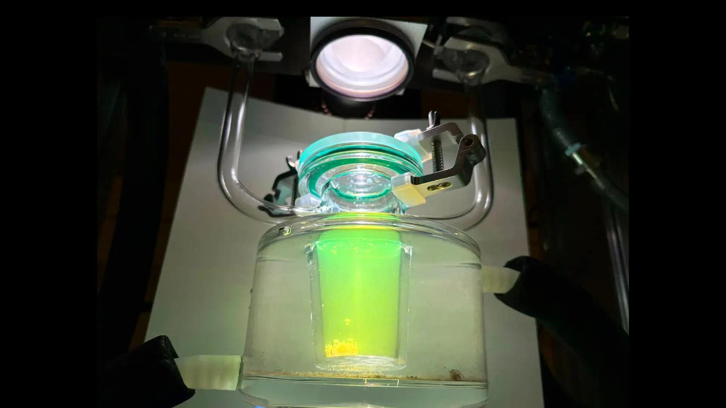
The new method sidesteps the problem by halting all cellular motion with extreme speed. The team designed a miniature freezing chamber that fits into an optical microscope. With an electrically triggered injection of cryogen, they can drop the temperature of the sample almost instantly. The freeze can be timed to within ±10 milliseconds of a chosen trigger, such as a laser flash or chemical pulse.
Related Stories
- New research finds that cells detect and react to sound waves
- Scientists recreate cellular metabolism within synthetic cells
“Instead of chasing speed in imaging, we decided to freeze the entire scene,” explained lead author Kosuke Tsuji. This shift in thinking turned the microscope into something like a time machine, able to capture a moving process at the exact instant of interest.
Once frozen, the sample is stable. Researchers can then take as long as needed to image it with high exposure times, producing crisp pictures with excellent signal-to-noise ratios. Chemical and physical states, such as ion concentrations and redox balance, are preserved far better than with traditional chemical fixatives.
Preserving What Chemicals Cannot
For decades, electron microscopists have used cryofixation to preserve cellular ultrastructure, since chemical treatments often distort delicate internal features. Under light microscopy, requirements are usually less strict. Yet freezing still offers striking advantages.
Chemical fixatives like formaldehyde can disrupt pH, damage fluorescent probes, or shift the distribution of ions. Freezing, by contrast, locks everything in place exactly as it was in life. This includes the folding of fluorescent probes, the position of calcium ions, or even subtle differences in molecular binding.
One of the study’s senior authors, Katsumasa Fujita, described the project as a bold rethinking of how to study cells: “This research began with a bold shift in perspective: to arrest dynamic cellular processes during optical imaging rather than struggle to track them in motion. We believe this will serve as a powerful foundational technique, offering new insights across life-science and medical research.”
From Calcium Signals to Frozen Waves
To show what their invention could do, the researchers tested it on one of biology’s fastest and most important signals: calcium ion waves. Calcium surges through cells to control heartbeats, muscle contractions, and nerve communication.
In their demonstration, the team triggered calcium waves in heart-muscle cells using ultraviolet light. Just as the wave swept across the cell, they froze it in place. When imaged later, the frozen wave could be studied in three dimensions using super-resolution methods that would normally be far too slow to follow the living wave in real time.
Masahito Yamanaka, another lead author, emphasized the strength of this approach: “Our technique preserves both spatial and temporal features of live cells with instantaneous freezing, making it possible to observe their states in detail. While cells are immobilized, we can take the opportunity to perform highly accurate quantitative measurements with a variety of optical microscopy tools.”
Accuracy Beyond the Limits of Live Imaging
With cells fixed in place, the researchers could use exposure times up to 1,000 times longer than possible during live imaging. This increased the measurement accuracy dramatically. In one test, cells loaded with fluorescent calcium indicators revealed exact distributions of both the ions and the probes themselves.
The method also allows different imaging techniques to be combined without timing issues. Normally, aligning separate methods like Raman spectroscopy and super-resolution fluorescence microscopy is challenging because each takes place over different timeframes. With frozen samples, however, multiple approaches can be applied in sequence to the same state of the cell, producing a layered view of its biology.
Precision on the Millisecond Scale
The ability to trigger freezing with 10-millisecond precision means researchers can choose which moment to capture with remarkable accuracy. That level of control is critical for events that last less than a second, such as protein-protein interactions or short bursts of signaling molecules.
In effect, the microscope now works like a synchronized high-speed camera and high-resolution still camera combined. The fast action is caught in real time, then held long enough for detailed study.
A New Tool for Cellular Discovery
By merging live imaging with cryogenic preservation, the technique opens a wide range of possibilities. Scientists can now:
- Capture fleeting molecular interactions too fast for normal observation.
- Study the movement of ions and molecules with both speed and precision.
- Improve the accuracy of super-resolution or 3D imaging without sacrificing clarity.
- Design new probes and sensors optimized for frozen conditions.
For many fields, from neuroscience to cancer biology, the ability to hold and inspect fast processes at leisure could be transformative.
Practical Implications of the Research
The ability to freeze living cells in action with millisecond accuracy could reshape biological research. For medical science, it provides a way to study fleeting signals in heart cells, neurons, or immune cells with clarity never before possible. This could reveal how diseases alter rapid signaling, from irregular heart rhythms to faulty nerve communication.
In drug development, the method offers a chance to capture molecular interactions at the instant they occur, helping to test how new treatments affect signaling pathways. It also creates opportunities for designing more reliable fluorescent probes and improving imaging technologies that already drive progress in medicine and biology.
By bridging speed and detail, this innovation could speed up discoveries across cell biology and lead to new treatments that rely on a deeper understanding of life at its fastest scales.
Note: The article above provided above by The Brighter Side of News.
Like these kind of feel good stories? Get The Brighter Side of News' newsletter.