Groundbreaking new technology detects near-invisible microplastics in the environment
In today’s world, microplastics – those minuscule particles smaller than 5 millimeters in diameter – are omnipresent.
[July 27, 2023: Staff Writer, The Brighter Side of News]
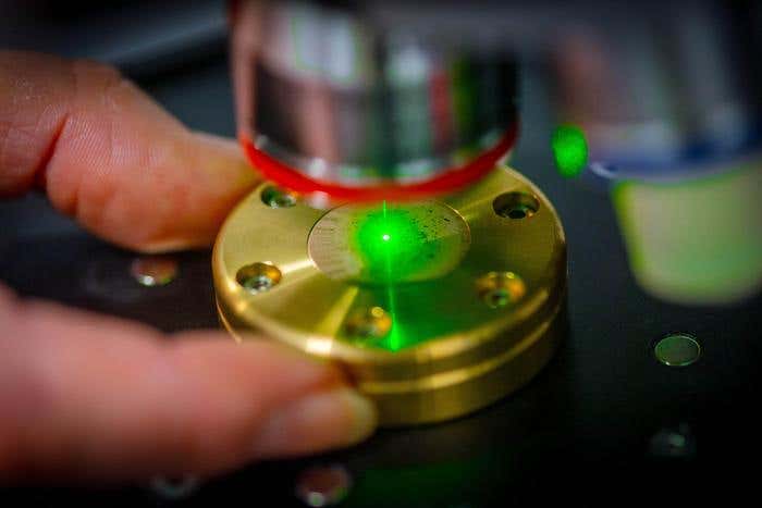
The researchers use a method known as Raman microspectroscopy. It works by shining a monochromatic laser source onto a sample and detecting the light scattered by the molecules. (CREDIT: Andreas Heddergott / TUM)
In today's world, microplastics – those minuscule particles smaller than 5 millimeters in diameter – are omnipresent. Whether you're sipping water, enjoying a meal, or simply breathing in a city, chances are you're interacting with them in some way. How much, though, do we really know about their concentration in the environment, our food, and our drinking water?
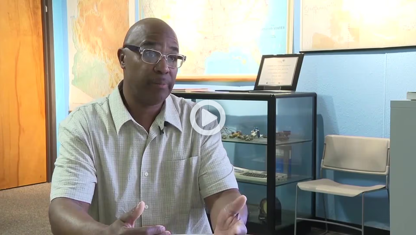
The Technical University of Munich (TUM) has recently made significant strides in unraveling this mystery. Spearheaded by Dr. Natalia Ivleva from the Chair of Analytical Chemistry and Water Chemistry at TUM, researchers have devised an innovative and automated method to both identify and quantify these particles.
The Microscopic Threat
These inconspicuous particles can absorb and transport harmful contaminants and toxins. Dr. Ivleva emphasizes, “We urgently need analytical techniques to learn about the size, concentration, and composition of these particles.”
Traditional methodologies have had their limitations. Analyzing the residue left after heating samples was the norm, but this posed problems. It failed to give insights into the number, size, and shape of these microplastic particles, making accurate quantification a challenge.
Related Stories
However, the TUM research initiative has redefined the approach.
“It is particle-based,” states Dr. Ivleva, drawing a distinction between their technique and older ones. Instead of obliterating the particles, the TUM method focuses on direct analysis. The secret weapon? Raman microspectroscopy.
At its core, this technique involves projecting a monochromatic laser onto a sample. The scattered light, when compared to the initial laser source, offers a wealth of information on the substance being examined. Specifically, for plastics like polyethylene, polystyrene, and polyvinyl chloride, this scattered light is as distinctive as a fingerprint, offering precise identification.
Revolutionizing Detection with Automation
The journey from ideation to execution was a meticulous one. Dr. Ivleva reminisces, “When we started, we still had to make manual measurements. It took us months to investigate a few thousand particles.”
Dr. Natalia Ivleva and her team have developed new methods for analyzing microplastics. (CREDIT: Andreas Heddergott / TUM)
The tediousness of such manual processes is a tale of the past now. Today, the team's software, the open-source TUM-Particle Typer 2, can churn out results in mere hours, making the identification of microplastics a seamless endeavor.
This software is a paragon of modern automation. Although particles still need to be sieved from their solutions and placed under the Raman microspectroscope, everything that follows is entirely automated. With a light microscope, the software can photograph, measure, and distinguish between particles and fibers. It then uses this data to calculate the number of particles and fibers, ensuring accurate results in the subsequent Raman spectroscopy.
Comparison of average detection rates between three experts, TUM-ParticleTyper (version 1) [14] and TUM-ParticleTyper 2 (this work) with Expert 1 being set as reference. (CREDIT: SpringerLink)
The Challenge of Nanoplastics
However, for particles smaller than 1 µm – termed nanoparticles – the detection process becomes trickier. These are almost invisible under a light microscope. To circumvent this, Dr. Ivleva and her team have employed a method involving size fractionation.
The tool of choice for this is a field flow fractionation (FFF) system. The FFF streamlines particles based on their size, effectively separating them. Combined with Raman spectroscopy, this specialized device facilitates the identification of various nanoplastics.
Graphical description of morphological parameters taken from each detected object. Additional useful features can be seamlessly integrated into this approach as it is generally meant as new suggestion to the topic of fiber recognition. (CREDIT: SpringerLink)
“The new analytical processes permit rapid and precise investigation of concentration, size, and composition of micro- and nanoplastics,” Dr. Ivleva proudly concludes. This landmark development promises a clearer understanding of these particles' influence on the environment and human health.
As microplastics continue to weave themselves intricately into our daily lives, it is tools like the one developed at TUM that will arm us with the knowledge to better comprehend and address this modern challenge.
For more green news stories check out our Green Impact section at The Brighter Side of News.
Note: Materials provided above by The Brighter Side of News. Content may be edited for style and length.
Like these kind of feel good stories? Get the Brighter Side of News' newsletter.