Scientists discover why long COVID affects lungs differently
New UVA study shows how immune cell changes in Long COVID patients reveal severity of lung damage and offer treatment hope.

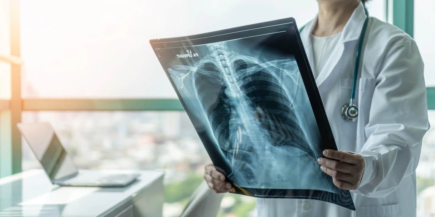
A major study from the University of Virginia finds that specific immune changes in Long COVID patients reveal the severity of their lung damage—offering new hope for targeted treatments. (CREDIT: Chinnapong / Shutterstock)
At first glance, some people seem to have recovered from COVID-19. They no longer test positive and can return to work or school. But for many, the virus leaves behind a lasting impact—especially in the lungs.
A groundbreaking new study from the University of Virginia School of Medicine and published in the journal, Nature Immunology, reveals why some people continue to struggle with breathing problems, even long after their infection.
The research shows that these lingering symptoms aren't random. Instead, they come from very specific changes in the immune system—changes that may be tied directly to how much damage COVID-19 caused in the lungs. The team studied 110 patients from UVA Health’s Long COVID Clinic, most of whom had severe COVID-19 and were hospitalized before vaccines were available.
The researchers found distinct “immune landscapes” based on lung condition. In people with milder damage, certain immune cells were active. In those with more severe lung scarring, other immune markers appeared. These discoveries open the door to more personalized treatments.
“Long COVID is complex, with a variety of potential underlying causes,” said Dr. Judith Woodfolk, one of the lead researchers. “Our findings reveal crucial differences in the blood that reflect the extent of lung damage.”
Immune Cells Tell the Story
One of the most exciting aspects of this study was its use of machine learning. The researchers trained a form of artificial intelligence to scan immune cell data and spot patterns. They looked at changes in T cells—key players in the immune system—and linked those changes to different types of lung disease.
People with less severe lung damage had large amounts of a type of T cell known as CCR5+CD95+CD8+ cells. These cells had specific features: they were active, often dividing, and seemed ready to fight infection. In contrast, patients with more severe damage had weaker T cell activity and high levels of a protein called CXCL13, which is known to encourage lung scarring.
Related Stories
The immune system looked very different depending on the type of lung disease. For example, people with restrictive lung disease—a condition where the lungs cannot expand fully—showed two main immune patterns. One group had more active T cells, while another had immune markers tied to scarring and worse outcomes.
Dr. Woodfolk explained, “By analyzing many different immune measures, we can pinpoint potential targets that may not only predict who might experience worse outcomes but also help guide more tailored and effective treatments in the future.”
Not All Breathlessness Is the Same
The team divided patients into five different groups based on lung function tests and walking ability. Some had severe scarring and low oxygen levels after walking. Others showed poor stamina but no lung restriction. A third group had abnormal blood pressure and heart rate responses after exercise, likely due to nervous system issues. These differences helped researchers understand how complex Long COVID really is.
Interestingly, 41% of patients improved over time, shifting into a group with milder lung problems. However, fibrotic lung damage—scarring of lung tissue—was still present in many even two years after their infection. In fact, only 13% of those who had lung imaging showed improvement in fibrosis scores.
Another surprise was that some patients had severe fatigue and breathing issues but normal lung function. Their problems may come from abnormal breathing patterns or issues with the nervous system rather than direct lung damage.
Still, the strongest signs of harm came from two specific groups—those with restrictive lung disease. Patients in the most severe group had lower oxygen levels, poor gas exchange, and high levels of fibrosis. Their immune systems showed clear signs of stress and inflammation.
T Cells Found Deep in the Lungs
To find out if the same immune cells found in blood were also in the lungs, researchers collected fluid samples from the airways of some patients. They found several T cell types there—cells that had the same markers as the ones seen in the blood. These included CD8+ T cells and CD4+ memory cells, both known for helping fight infection and creating long-term immunity.
Many of these lung T cells also had markers that help them stay in tissue, such as CD69 and CD103. This means the cells are not just passing through—they’re staying in the lungs long after infection and may be influencing recovery or ongoing symptoms.
The researchers also found that some of these T cells were virus-specific, reacting to SARS-CoV-2 proteins like the spike and nucleocapsid. These cells made inflammatory molecules like interferon-gamma (IFNγ), interleukin-2 (IL-2), and tumor necrosis factor (TNF), which can be useful during infection but harmful if produced for too long.
When the team looked closer, they saw that certain CD8+ T cells were more common in patients with severe lung damage. These cells were in a state that suggested long-term activation. In some cases, the more of these cells a person had, the worse their lung function was.
“By uncovering distinct immune patterns in patients who have different types of restrictive lung disease after infection, we can better understand the immune drivers of lung injury,” said Dr. Woodfolk.
Toward Better Treatment and Diagnosis
The team didn’t just study T cells. They also measured 76 different plasma proteins, 73 types of autoantibodies, and over 100 immune cell types. One finding stood out: patients with the most scarring had much higher levels of CXCL13. This molecule is linked to fibrosis and has been seen in other lung diseases.
Other immune signals also popped up. These included type 1 inflammation markers like CXCL10 and TNF, as well as indicators of an overactive immune response such as IL-6 and IL-1RA. These markers may help doctors spot patients at risk of worsening lung function.
Using advanced data analysis, researchers created models that could separate patients with severe lung issues from healthy people just by looking at immune markers. This approach could lead to better tools for diagnosing Long COVID and deciding who might need more aggressive treatment.
Dr. Woodfolk shared her hope: “Our ultimate goal is to help patients by guiding new treatments that could stop or even reverse lung damage caused by COVID-19.”
While these findings are early, they give scientists a better map of how Long COVID affects the immune system and the lungs. By continuing to explore these immune pathways, researchers may eventually find ways to heal or even prevent the long-term damage that COVID-19 can cause.
Note: The article above provided above by The Brighter Side of News.
Like these kind of feel good stories? Get The Brighter Side of News' newsletter.


