Smell and sight can spot Parkinson’s disease years earlier, study finds
New brain scan study finds changes in smell and sight responses may detect Parkinson’s years before symptoms start.
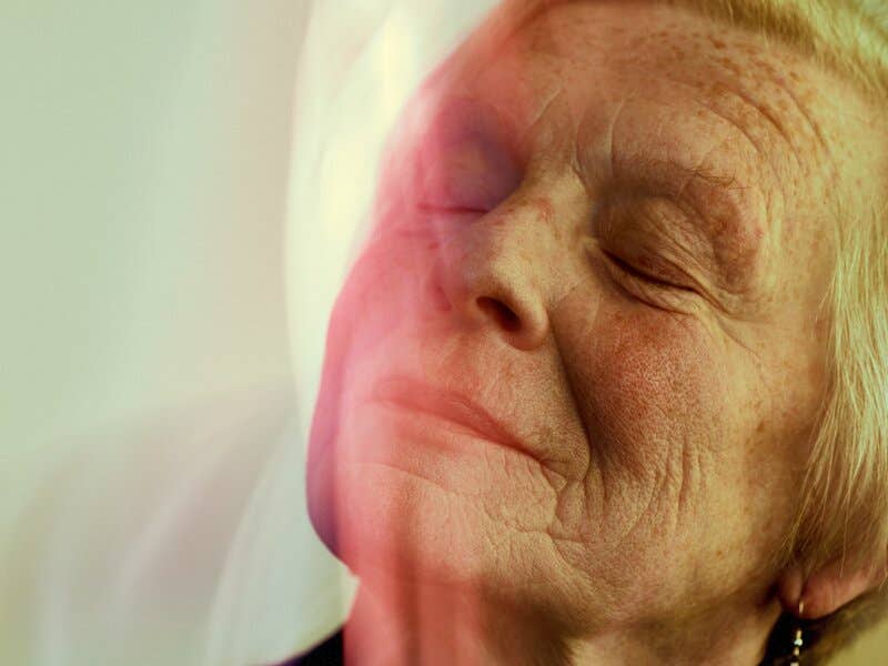
A new study using advanced brain scans shows how changes in smell and sight could offer early signs of Parkinson’s disease. (CREDIT: CC BY-SA 4.0)
Diagnosing Parkinson's disease has always been a race against time. The later it’s found, the harder it is to treat. But a group of researchers may have just unlocked a way to detect this illness earlier than ever before. Their new method focuses on how the brain processes two simple senses: smell and sight.
Scientists from the Champalimaud Foundation in Portugal, working with teams from the University Medical Center Gottingen, believe they’ve found a possible early warning sign for Parkinson’s. Published in the Journal of Cerebral Blood Flow and Metabolism, their study used high-resolution brain scans in mice to identify patterns of brain activity that could help predict the disease years before symptoms show.
Why the Senses Matter in Parkinson's
Parkinson’s disease is known for shaking hands, stiff muscles, and slow movement. But what many don’t realize is that it often starts quietly. People can begin to lose their sense of smell five to ten years before any tremors begin. They may also develop visual problems, even hallucinations. Still, these symptoms alone aren’t enough to diagnose the disease.
The problem is that losing your sense of smell or sight doesn't always mean you have Parkinson’s. Many people experience these issues and never develop the disease. That’s why scientists have been looking for a more reliable way to spot Parkinson’s earlier.
Noam Shemesh, who leads the Preclinical MRI lab at the Champalimaud Foundation, wanted to go further. Along with Tiago Outeiro, a Parkinson’s specialist in Germany, they tried something new: testing both senses at the same time using ultra-high-resolution brain scans. “The vast majority of fMRI studies in animal models focus on a single sense,” Shemesh said. “We analyzed both visual and olfactory sensory modalities. That's pretty rare.”
Powerful Imaging, Clearer Answers
To test their idea, the researchers used a powerful type of brain scanner called fMRI — functional magnetic resonance imaging. This tool lets scientists see which parts of the brain are active in real time by tracking blood flow and oxygen levels.
Related Stories
Their scanner was no ordinary machine. It produced a magnetic field of 9.4 Tesla, three times stronger than most hospital machines. This allowed them to study the brains of genetically engineered mice that produce too much of a human protein called alpha-synuclein. That protein builds up in the brains of people with Parkinson’s and is believed to trigger the disease.
When these mice were shown light or exposed to smells, the researchers watched how their brains responded. Compared to healthy mice, the Parkinsonian mice showed far less brain activity in areas responsible for processing those senses. Francisca Fernandes, one of the lead analysts, found that this lack of response was consistent across both senses.
But the team knew fMRI data could be tricky. Brain activity seen in scans depends not just on neurons but also on how well blood vessels work. Shemesh explains, “It doesn't detect neural activity per se. Since it relies on interactions between ongoing neural activity and vascular properties, it detects a complicated combination of both effects.”
So, to separate those effects, the team added two more tests. One measured blood flow using a method called pseudo-continuous arterial spin labeling (pCASL). The other looked for a protein called C-FOS, which appears when neurons fire. These tests confirmed that the mice had both reduced blood flow and less neural firing, with the neurons showing more severe decline.
A Real Path Toward Early Detection
The most exciting part of the study is what it suggests for future human testing. If similar brain activity patterns can be seen in people who are just beginning to lose their sense of smell or vision, doctors may have a powerful new tool. Early detection could mean early treatment, which might slow the disease before it causes major damage.
Shemesh sees promise: “If we saw something weird in both sensory modalities, we could potentially say that there is something more global happening in their neural circuits, and that we need to follow up on that.”
Because fMRI is non-invasive and widely used, this method could be added to current screening tools with little risk. Outeiro agrees: “This could add to the toolbox for diagnosing and classifying PD, something that is urgently needed.”
Importantly, this study is the first to show these specific brain activity patterns across both smell and vision in a well-studied mouse model. These mice overproduce alpha-synuclein, just like humans with Parkinson’s. They also start to show movement problems at a similar age in mouse years — around nine months — when the scans were taken.
More Than Movement: The Broader Impact of PD
Parkinson’s is not just a movement disorder. It causes serious problems in the brain’s communication systems. Many of these begin before visible symptoms appear. As alpha-synuclein builds up, it forms harmful clumps known as Lewy bodies. These disrupt how neurons work, especially in a region called the substantia nigra, which makes dopamine. Losing dopamine leads to motor issues like tremors and stiffness.
But even before that damage, changes happen in brain areas tied to smell and sight. Studies have shown that mice overproducing alpha-synuclein lose their ability to recognize smells and see clearly. In some mice, the retina — the light-sensing part of the eye — becomes thinner and stops working properly. Their brain activity during smell and light tests drops sharply.
By using c-FOS protein levels as a guide, this study showed that brain activity fell by over 50% in Parkinsonian mice. Meanwhile, their blood flow dropped by about 10%. This suggests that the main problem lies in the neurons themselves, not just poor circulation.
Outeiro emphasized how useful this mouse model is: “It produces the human type of alpha-synuclein.” That makes it an excellent model for testing both new diagnostic methods and potential treatments.
A Hopeful Future for Earlier Treatment
For now, this discovery is still in its early stages. But the findings open the door to new research. If doctors can spot Parkinson’s before it takes hold, they might have a better chance at stopping it or at least slowing it down.
The Champalimaud team is already looking ahead. They hope future studies will check if the same brain changes can be seen in humans. If people complaining of smell loss or visual problems also show these fMRI patterns, early treatment may become a reality.
Shemesh is optimistic: “It gives us some hope that, with future studies, there will be more things we can look at that hint at early development stages of PD and also determine which treatments might help if they're given early on.”
The research was supported by the €200,000 Mantero Belard Award, given by Santa Casa da Misericórdia de Lisboa. That support helped fund the state-of-the-art equipment used in this study.
Early signs like changes in how the brain smells and sees might one day become powerful tools for fighting one of the world’s most common brain disorders. For the millions who live with Parkinson’s — and for those who may develop it in the future — that’s a hopeful step forward.
Note: The article above provided above by The Brighter Side of News.
Like these kind of feel good stories? Get The Brighter Side of News' newsletter.