Major breakthrough reveals how cells create their cilia
Scientists identify SP5 and SP8 as key genetic switches that trigger primary cilia formation and may help explain ciliopathies.
 MSKCC Writer: Nick Gardner

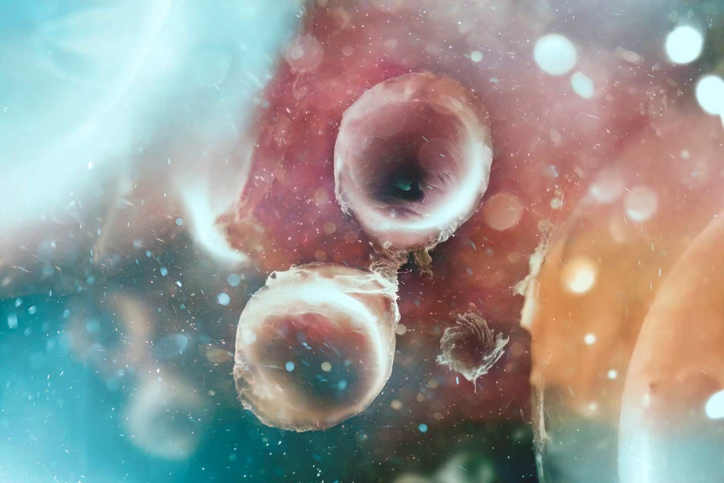
Researchers at Memorial Sloan Kettering Cancer Center have identified SP5 and SP8 as master genetic switches that trigger primary cilia formation, solving a major mystery in developmental biology and opening new avenues for studying ciliopathies. (CREDIT: Shutterstock)
- SP5 and SP8 help activate the genetic program that builds primary cilia, the signaling projections on many cells.
- Removing both genes reduced cilia formation in mouse embryos and stem-cell models, while adding SP8 induced cilia in normally unciliated cells.
- The findings clarify a developmental mechanism but do not demonstrate a treatment for human ciliopathies.
Cells that normally lack a primary cilium began growing one when researchers increased the activity of a single gene. The experiment helped identify two regulatory proteins, SP5 and SP8, that coordinate the construction of these tiny signaling structures. Their discovery addresses a basic question: what tells a cell to build its antenna?
The work, led by Yinwen Liang and Alexandra Joyner at Memorial Sloan Kettering Cancer Center, appeared in Science. It combined comparisons of mouse embryonic tissues with experiments in stem cells and developing mice. The results connect gene regulation with the formation of an organelle important for development and health.
They also establish important boundaries for interpreting the discovery. Cilia became fewer and shorter when both genes were removed, but did not disappear completely. Adding SP8 could initiate primary cilia in the tested settings, rather than establishing a universal way to rebuild every type of cilium.
Why cells need a signaling antenna
A primary cilium is a slender projection extending from a cell’s surface. Unlike the beating cilia that help move mucus through airways, it generally does not move. Instead, it organizes signals that help a cell respond to its surroundings.
Those signals have major consequences during development. Work involving the late MSK scientist Kathryn Anderson established an important connection between cilia and Hedgehog signaling in 2003. Hedgehog proteins provide messages that help organize developing tissues, including the neural tube that gives rise to the brain and spinal cord.
Defects in cilia can disrupt this communication and contribute to disorders collectively called ciliopathies. Depending on the affected genes and tissues, these conditions can involve the kidneys, brain, sensory systems or organ development. Understanding how cilia form therefore concerns much more than the appearance of a small cellular structure.
Researchers already knew many proteins required to construct or maintain cilia. What remained less clear was how cells coordinate the production of those components. Liang and Joyner investigated whether a small number of transcription factors, proteins that regulate gene activity, might help initiate that program.
Comparing cells with different building instructions
Liang initially examined whether cells without cilia were actively dismantling them. Experiments intended to block that disassembly had little effect on whether the structures appeared. The result encouraged the team to investigate instructions for construction rather than focusing only on destruction.
Mouse development offered a useful comparison. Many embryonic cells possess primary cilia, while a particular layer called the yolk sac visceral endoderm normally lacks them. Comparing these populations could reveal regulatory differences associated with the decision to build a cilium.
The team used single-cell RNA sequencing to measure which genes were active in individual cells. More than 100 cilia-related genes showed higher activity in ciliated populations. That pattern suggested coordinated regulation, rather than a change affecting just one structural component.
A complementary technique, ATAC-seq, identified accessible regions of DNA that regulatory proteins could potentially reach. Accessibility provides clues about gene regulation, but does not by itself prove that a gene is active. Combining these observations helped identify SP5 and SP8 as candidates for controlling the wider cilia-building program.
Removing the regulators, then adding one back
The researchers tested those candidates through genetic manipulation. In gastruloids, stem-cell structures that model aspects of early embryonic development, removing both genes reduced the proportion of ciliated cells. The measured fraction fell from 48.5% in controls to 23.8% in the double-knockout models.
The remaining cilia were also shorter. Average length fell from 1.3 micrometers in controls to 0.9 micrometers in the modified models. A micrometer is one-millionth of a meter, underscoring the small scale at which the regulatory changes affected cell structure.
Related experiments in mouse embryos found reductions in cilia number and length across multiple tissues. These observations strengthened the case that the genes contribute to normal cilia formation beyond a single culture condition. Residual cilia also showed that removing the genes did not uniformly abolish the structure.
The reverse experiment provided another important test. Introducing additional SP8 into normally unciliated extraembryonic cells caused many to form primary cilia. “If you add SP8 to extraembryonic cells, many of the cells now make cilia,” Joyner said.
Activating a network rather than one component
SP5 and SP8 are transcription factors rather than the structural material from which a cilium is assembled. Their importance lies in regulating other genes. The team found evidence that the proteins bind regulatory regions associated with numerous cilia-related genes and help maintain their expression.
Their targets include genes involved in the organelle’s construction and other transcription factors associated with ciliary programs. This arrangement places SP5 and SP8 upstream of several parts of the process. It helps explain how altering two regulators could affect a complex structure built from many components.
Coordinating many genes matters because a cilium needs more than one kind of cellular machinery. Its framework must be assembled, proteins must reach the right location, and the structure must retain the components used for signaling. An upstream regulator can influence several of these requirements at once, giving researchers a way to study how their coordination succeeds or fails.
The results do not make DNA binding and gene activation interchangeable findings. Binding identifies a potential direct regulatory connection, while changes in gene expression provide evidence about its consequences. Genetic loss and gain experiments add another layer by testing whether those regulatory changes accompany altered cilia formation.
“We see it as a big breakthrough to find the upstream transcription factors that switch the whole thing on,” Liang said. The experiments support a coordinating role for SP5 and SP8 in the systems studied. Whether the same control operates identically in other tissues or developmental stages remains a separate question.
Developmental consequences and unanswered questions
The mutant mice also showed abnormalities involving motile cilia. At the embryonic node, these moving structures help establish the body’s left-right organization. Defects there can disrupt organ placement, including producing situs inversus, in which internal organs occupy reversed positions.
Hydrocephalus, an abnormal accumulation of fluid in the brain, also occurred in mutant animals. Such findings connect the regulators with developmental problems resembling features of human ciliopathies. They do not establish that mutations in SP5 or SP8 account for those disorders in patients.
Importantly, inducing a primary cilium is different from creating a working array of motile cilia. Increasing SP8 did not establish a general method for producing motile cilia in the tested systems. The genetic instructions and cellular conditions required for the different structures therefore cannot be treated as identical.
The work provides a starting point for building better experimental models and investigating candidate disease genes. No treatment restoring cilia in patients was tested, and activating a regulator cannot be assumed to repair a defective structural protein. A key next step is determining where these regulators can improve cilia formation and whether the resulting structures support normal signaling.
Dig deeper into cilia formation and disease
These resources explain ciliary gene regulation, assembly, signaling and the disorders caused by disrupted function.
Specification of Ciliated Cells
This review places cilia-building transcription factors within the broader signals that determine which cells develop different types of cilia. (Annual Review of Cell and Developmental Biology, April 2026)
The intraflagellar transport cycle
This review explains the transport machinery that supplies and maintains cilia after the cellular building program begins. (Nature Reviews Molecular Cell Biology, November 2024)
Cellular signalling by primary cilia in development, organ function and disease
This review describes how cilia organize signaling pathways and why disrupted communication can affect multiple organs. (Nature Reviews Nephrology, February 2019)
Genes and molecular pathways underpinning ciliopathies
This review connects disease-associated genes with different parts of ciliary structure and function, explaining the diversity of ciliopathies. (Nature Reviews Molecular Cell Biology, July 2017)
Hedgehog signalling in the mouse requires intraflagellar transport proteins
This mouse study helped establish the connection between ciliary transport machinery and Hedgehog signals during embryonic development. (Nature, November 2003)
Research findings are available online in the journal Science.
The original story "Major breakthrough reveals how cells create their cilia" is published in The Brighter Side of News.
Related Stories
- Engineered cells deliver healing signals directly into wounds
- Scientists build synthetic cells with programmable DNA pores
- Blood cells originated from single-celled ancestors 700 million years ago
Like these kind of feel good stories? Get The Brighter Side of News' newsletter.
Rebecca Shavit
Writer
Based in Los Angeles, Rebecca Shavit is a dedicated science and technology journalist who writes for The Brighter Side of News, an online publication committed to highlighting positive and transformative stories from around the world. Having published articles on MSN, AOL News, and Yahoo News, Rebecca's reporting spans a wide range of topics, from cutting-edge medical breakthroughs to historical discoveries and innovations. With a keen ability to translate complex concepts into engaging and accessible stories, she makes science and innovation relatable to a broad audience.