Scientists develop 3D-printed ceramic bone implants — just like real human bone
Researchers created 3D-printed ceramic bone implants that closely mimic human bone and support tissue regeneration.

 Edited By: Joseph Shavit
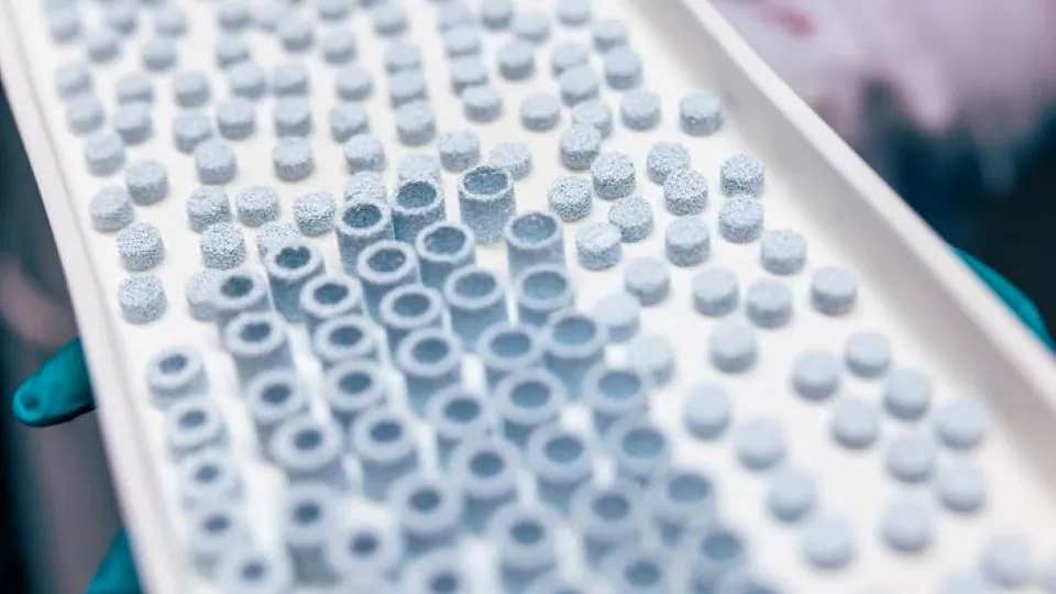
Finnish researchers developed personalized 3D-printed ceramic bone implants that imitate natural bone and support healing. (CREDIT: Jonne Renvall, Tampere University)
Millions of people each year undergo painful procedures to repair damaged or missing bone. Whether caused by injury, aging or disease, bone loss can dramatically affect mobility, independence and quality of life. Now, researchers at Tampere University in Finland say they may have developed a more personalized and accessible solution using 3D-printed ceramic implants designed to closely imitate natural human bone.
The research team created bone-like scaffolds using hydroxyapatite, the same mineral compound found in real bone tissue. By combining this material with advanced ceramic 3D printing, the scientists produced implants with carefully controlled internal structures that support the body’s natural ability to rebuild bone.
The findings could mark a major step toward customized bone implants designed specifically for each patient. Researchers believe the technology may eventually replace some traditional bone graft procedures, which often require tissue from donors or the patient’s own body.
“By using the same material that nature uses and shaping it through ceramic 3D printing, the implants can be precisely tailored to match a patient’s individual bone defect, without relying on drugs or growth factors that may cause side effects,” said Antonia Ressler, Postdoctoral Research Fellow at the Tampere Institute for Advanced Study.
A Growing Need For Better Bone Repair
Bone grafting remains one of the world’s most common transplant procedures. More than 2 million operations are performed annually. Current treatments often depend on bone harvested from another part of the patient’s body or from donated tissue.
These procedures can create additional pain and complications. Patients may face bleeding, nerve damage, long recovery periods and limited donor tissue availability. As populations continue aging worldwide, the demand for safer and more effective bone regeneration methods is rapidly increasing.
The global market for bone graft materials continues expanding as doctors search for alternatives that heal faster and reduce complications. Scientists have long tried to create synthetic scaffolds that imitate natural bone while supporting tissue growth inside the body.
Natural bone itself is highly complex. Trabecular bone, the spongy inner structure found in many bones, contains interconnected pores that allow nutrients, cells and blood vessels to move throughout the tissue. Replicating that structure artificially has proven difficult.
Printing Bone Layer By Layer
The Tampere University researchers used an advanced manufacturing process called ceramic vat photopolymerization. The method allows scientists to build ceramic structures layer by layer with extremely high precision.
In the process, a light-sensitive resin mixed with ceramic particles hardens when exposed to laser light. Each layer measures only about 25 micrometers thick, thinner than a human hair. After printing, the scaffold undergoes high-temperature processing called sintering, which strengthens the material.
This approach gave researchers remarkable control over pore size, wall thickness and internal connectivity. Those details matter because cells need enough space to enter the scaffold, communicate and form new tissue.
Using micro-computed tomography scans, the researchers tested four different scaffold designs with varying porosity and pore sizes. They discovered that scaffolds containing pores around 400 micrometers wide and about 45% porosity created the best balance between strength and biological performance.
“This architecture achieved a crucial balance between strength and biological performance, allowing bone-forming cells to enter the material, interact with one another, and successfully begin forming new bone tissue,” Ressler said.
Finding The Right Balance Between Strength And Healing
Creating an ideal bone scaffold involves balancing competing needs. The structure must remain strong enough to support damaged bone while also allowing living cells to grow inside it.
The team tested hydroxyapatite discs processed at temperatures between 900 and 1300 degrees Celsius. Human osteoclasts, cells responsible for breaking down and remodeling bone, were then placed onto the surfaces.
The results showed that lower processing temperatures preserved better biological activity. Cells attached strongly to scaffolds processed at 900, 1000, 1100 and 1200 degrees Celsius. However, no cells successfully attached at 1300 degrees Celsius.
Researchers found that increasing temperature improved mechanical strength but reduced surface properties needed for healthy cell growth. At extremely high temperatures, the scaffold surface became less welcoming to human cells.
“We found that the high temperatures required during processing can alter the surface of the material in ways that make it more difficult for human cells to attach,” Ressler explained. “Our finding highlights that not only the composition, but also the surface properties of biomaterials are critical for successful bone regeneration.”
Ultimately, the team selected 1000 degrees Celsius as the optimal processing temperature because it balanced strength and biological compatibility.
How Human Cells Responded
To evaluate healing potential, scientists seeded human bone marrow stem cells onto the scaffolds. These stem cells can transform into osteoblasts, the cells responsible for building bone.
The researchers observed strong cell survival across all scaffold types. However, the most successful bone formation occurred in scaffolds with moderate pore sizes and carefully balanced porosity.
One scaffold design, called HAp2, showed especially promising results. It had pores averaging around 400 micrometers and a porosity level near 45.6%.
Cells growing inside HAp2 produced stronger collagen networks and higher levels of osteocalcin, a protein tied to mature bone formation. The scaffold also maintained acceptable compressive strength within the lower range of natural trabecular bone.
By contrast, scaffolds with extremely large pores supported more cell growth initially but struggled to create organized bone tissue. Researchers found that simply increasing pore space did not guarantee better healing.
Trace Elements Produced Unexpected Challenges
The team also experimented with adding trace elements such as magnesium, zinc and strontium into the hydroxyapatite scaffolds. These minerals are known to support bone growth in certain settings.
However, the high temperatures used during manufacturing altered the material’s chemistry. Part of the hydroxyapatite transformed into another calcium phosphate phase called beta-tricalcium phosphate.
This chemical shift changed the surface charge and wettability of the scaffolds. Surfaces became more hydrophobic, meaning they repelled water more strongly. That made it harder for cells to attach and grow.
Cell growth declined as trace element concentration increased. The highest-substituted scaffolds showed the weakest osteoclast formation and little evidence of normal bone remodeling.
The findings revealed how sensitive human cells are to tiny changes in surface chemistry. Even beneficial minerals can create problems if manufacturing conditions alter the final material structure.
Personalized Bone Implants May Arrive Within A Decade
The researchers believe their work lays an important foundation for future personalized medicine. Because ceramic 3D printing allows computer-guided customization, implants could eventually be designed to match each patient’s exact bone defect.
“This technology allows implants to be designed for individual needs, no more ‘one size fits all’ solutions,” Ressler said. “We believe these types of implants could be used in routine bone regeneration treatments within the next decade.”
The research emerged from the AffordBoneS project funded through the Horizon Europe Marie Skłodowska-Curie Postdoctoral Fellowship programme. A follow-up project called GlassBoneS aims to continue developing affordable bone scaffolds for wider clinical use.
Practical Implications Of The Research
This technology could eventually reduce the need for painful bone harvesting procedures from patients or donors. Personalized ceramic implants may shorten recovery times, lower surgical complications and improve long-term healing outcomes.
The research also provides valuable design guidelines for future bone regeneration materials. Scientists now have stronger evidence that pore sizes near 400 micrometers and porosity around 45% create an effective balance between structural strength and biological performance.
Beyond orthopedics, the findings may influence regenerative medicine more broadly. Understanding how surface chemistry, porosity and material processing affect cell behavior could help researchers improve implants for dental repair, spinal injuries and other tissue engineering applications.
If future clinical studies succeed, 3D-printed ceramic implants may become more affordable and accessible worldwide, offering millions of patients safer and more personalized treatment options.
Research findings are available online in the journal Materials Today Bio.
The original story "Scientists develop 3D-printed ceramic bone implants — just like real human bone" is published in The Brighter Side of News.
Related Stories
- Breakthrough drug-delivery patch promotes regeneration of heart tissue
- First-of-its-kind drug promotes organ and heart tissue regeneration
- New electronic skin heals and adapts like human tissue
Like these kind of feel good stories? Get The Brighter Side of News' newsletter.
Hannah Shavit-Weiner
Medical & Health Writer
Hannah Shavit-Weiner is a Los Angeles–based medical and health journalist for The Brighter Side of News, an online publication focused on uplifting, transformative stories from around the globe. Having published articles on AOL.com, MSN and Yahoo News, Hannah covers a broad spectrum of topics—from medical breakthroughs and health information to animal science. With a talent for making complex science clear and compelling, she connects readers to the advancements shaping a brighter, more hopeful future.