Why do we get sleepy? New study reveals brain cells that help drive sleep
Sleep drive neurons in mice may help the brain track wakefulness and trigger deeper recovery sleep after long hours awake.
 University of Basel Writer: Angelika Jacobs

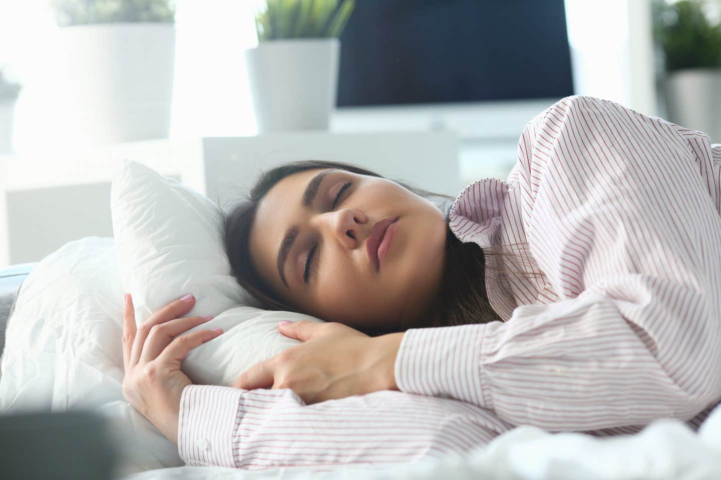
Mice slept far less when key brainstem cells were blocked, giving researchers a clearer look at how sleep pressure builds. (CREDIT: Shutterstock)
- Certain brain cells in mice became more active the longer the animals stayed awake, then quieted after sleep began.
- Turning on those cells made rested mice sleep longer and more deeply, while blocking them sharply reduced sleep.
- The findings give sleep researchers a clearer way to study how the brain builds the pressure to sleep.
Sleep pressure has a stubborn way of winning. After enough time awake, alertness starts to slip, and the need for sleep can feel less like a preference than a command.
That pressure, often called sleep drive, helps explain why a long day or sleepless night leads to deeper and longer recovery sleep. Yet a basic question has remained difficult to answer: how does the brain track wakefulness and turn it into the need to sleep?
A research team led by Professor Alex Schier at the Biozentrum, University of Basel, in collaboration with Beth Israel Deaconess Medical Center and Auburn University, has identified specific neuron groups in mice that help answer that question. The cells became active during prolonged wakefulness and helped promote sleep afterward.
“We have identified neuronal populations that monitor prolonged wakefulness and actively promote sleep,” says Alex Schier. “This is an important missing piece of the puzzle in understanding why we become sleepy.”
Brain cells that rise with wakefulness
The team compared brain activation in mice during normal sleep-wake cycles, sleep deprivation and recovery sleep. That approach pointed to brain areas whose activity appeared to reflect time spent awake.
Two areas stood out: the anterior medial preoptic area and the median raphe. The median raphe sits in the brainstem, while the anterior medial preoptic area lies in a region linked to sleep regulation. Within these areas, the researchers focused on neuronal populations that became more active as mice stayed awake.
In the median raphe, two cell types drew special attention: GABAergic neurons and serotonergic neurons. GABAergic neurons use a chemical signal linked to inhibition in the brain. Serotonergic neurons use serotonin, a chemical messenger involved in many brain functions.
The activation of both populations rose the longer the animals stayed awake. After sleep began, their activation declined again.
Testing whether the cells cause sleep
The next question mattered more than the map itself. Were these neurons merely recording that an animal had stayed awake, or were they helping create the push toward sleep?
To test that, the researchers artificially activated cells that had responded to sleep deprivation. When they activated deprivation-responsive cells in the median raphe or anterior medial preoptic area, mice slept longer and more deeply. The sleep resembled the recovery sleep that usually follows prolonged wakefulness.
Inhibiting those same cells produced the opposite result. Mice slept less and maintained alert wakefulness for longer periods.
“These neurons do not simply signal that an animal has been awake,” says Schier. “Our experiments show that they are crucial to promote sleep, and that they may be key components of the neural circuitry that generates sleep drive.”
The researchers tracked sleep using EEG recordings, which measure electrical activity in the brain. These recordings helped distinguish wakefulness, non-rapid eye movement sleep and rapid eye movement sleep.
The effects were strongest for non-rapid eye movement sleep, often shortened to NREM sleep. That stage is closely tied to recovery after sleep loss. Activating the cells also increased slow-wave activity in the delta range, a marker of sleep intensity that normally rises after deprivation.
Not every wake-active neuron pushed toward rest
The broader brain map showed that wakefulness activates many different regions. Some activity appeared early during sleep deprivation, then faded. Other activity peaked during recovery sleep. The pattern most relevant to sleep pressure built up during wakefulness and declined after sleep.
The researchers used whole-brain activity mapping across 162 brains and 26 conditions. In some experiments, mice were kept awake for six hours during the light phase, when they normally rest. The team used light water misting or new objects in the cage to keep them awake.
Those details mattered because they helped separate true sleep-pressure signals from responses to stimulation or novelty. The median raphe and anterior medial preoptic area showed activity patterns that tracked time awake in both forced and natural sleep-wake conditions.
But the study also found a cautionary twist. Some cells that became active during wakefulness did not promote sleep. In the lateral hypothalamic area, deprivation-responsive cells strongly promoted wakefulness when activated.
Those cells expressed orexin, a molecule known for helping maintain wakefulness. That finding showed that wake-activated cells can serve different roles. Some help keep an animal awake. Others help drive it toward sleep.
A sharp reduction in sleep drive
The most striking experiment came when the researchers inhibited both GABAergic and serotonergic neurons in the median raphe over a longer period. Mice with those cells suppressed slept approximately 70% less than usual.
They also stayed awake more than 6.5 hours longer each day than control mice. Their average wake bouts became more than twice as long. The sleep reduction lasted across several weeks.
The manipulation carried risks. About 17% of the mice died, likely because their sleep was reduced even more severely. Yet most survived. Unexpectedly, many did not show some of the severe behavioral impairments that usually accompany sleep deprivation.
They did not show the same strong buildup of sleep pressure seen in control mice. They also made no sleep attempts during deprivation and did not show significant rebound sleep afterward, despite sleeping far less overall.
The team also tested behavior. The sleep-reduced mice moved more, but they did not show significantly increased anxiety-like behavior in open field or elevated plus maze tests. They also remained behaviorally engaged with wood blocks placed in their cages.
In a contextual fear-conditioning task, the mice could still form long-lasting memories. Their memory appeared modestly weaker after one day, but by 14 days it reached levels comparable with controls.
A circuit with more than one switch
Further work showed that the median raphe contains sleep-promoting and wake-promoting cell types. GABAergic and serotonergic cells promoted NREM sleep and stronger delta power. Activating both together produced longer-lasting sleep than activating either group alone.
Glutamatergic cells in the same region worked differently. When the researchers activated those cells, mice became more awake and NREM sleep dropped sharply. Inhibiting them increased NREM sleep during the dark phase.
The researchers also found that sleep deprivation changed the internal properties of median raphe GABAergic cells. After six hours awake, these cells became more excitable. Their firing threshold dropped, and more of them fired spontaneous action potentials.
Serotonergic cells did not show the same significant shifts in resting membrane potential or firing threshold. Still, together with GABAergic cells, they helped produce sleep that looked like the intense recovery sleep after deprivation.
“Future studies could reveal how these neurons interact with the rest of the brain, and how sleep drive is generated at the molecular level,” says Dr. William Joo, first author of the study. “Our ability to stably transform sleep behavior also allows us to explore adaptations to long-term sleep loss – this may eventually reveal ways to confer resilience to sleep deprivation and other physiological challenges.”
Dig deeper into sleep pressure and the brain circuits that regulate rest
These resources explore how neural circuits, molecular changes and circadian rhythms influence sleep need and recovery after prolonged wakefulness.
The systems biology of sleep: toward integrative understanding of molecular and circuit-based mechanisms of sleep: Reviews how molecular pathways and neural circuits work together to regulate sleep. (Current Opinion in Biotechnology, 2026)
Revisiting brain gene expression changes and protein modifications tracking homeostatic sleep pressure: Examines molecular markers of accumulated sleep need and how their responses vary across brain regions and cell types. (npj Biological Timing and Sleep, 2025)
Prefrontal synaptic regulation of homeostatic sleep pressure revealed through synaptic chemogenetics: Shows how experimentally strengthening prefrontal connections in mice increases NREM sleep and delta-wave activity. (Science, 2024)
Sleep and circadian rhythmicity as entangled processes serving homeostasis: Explores the interaction between sleep regulation and daily biological rhythms. (Nature Reviews Neuroscience, 2024)
Sleep need, the key regulator of sleep homeostasis, is indicated and controlled by phosphorylation of threonine 221 in salt-inducible kinase 3: Investigates a protein modification that helps indicate and regulate sleep need in mice. (Genetics, 2023)
Research findings are available online in the journal Nature.
The original story "Why do we get sleepy? New study reveals brain cells that help drive sleep" is published in The Brighter Side of News.
Related Stories
- Melatonin may ease chronic pain while improving sleep, study finds
- Scientists reveal what caffeine actually does to your sleep
- Too much and too little sleep linked to faster aging
Like these kind of feel good stories? Get The Brighter Side of News' newsletter.
Rebecca Shavit
Writer
Based in Los Angeles, Rebecca Shavit is a dedicated science and technology journalist who writes for The Brighter Side of News, an online publication committed to highlighting positive and transformative stories from around the world. Having published articles on MSN, AOL News, and Yahoo News, Rebecca's reporting spans a wide range of topics, from cutting-edge medical breakthroughs to historical discoveries and innovations. With a keen ability to translate complex concepts into engaging and accessible stories, she makes science and innovation relatable to a broad audience.