Ancient viral DNA offers clues to why human and chimpanzee brains evolved differently
Lund researchers linked ancient viral DNA to a silenced chimpanzee gene, while human organoid tests revealed a role in developing nerve-cell extensions.
 Lund University Writer: Anna Elizabeth Hellgren

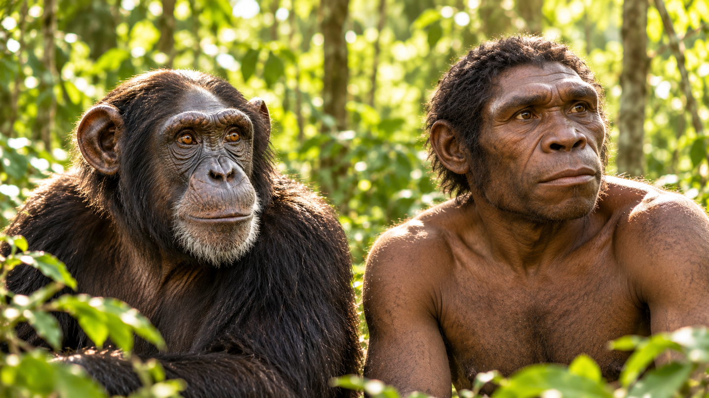
Ancient viral DNA silences LINC00662 in chimpanzee cells. Human organoid tests link the gene to early nerve-cell maturation. (CREDIT: Shutterstock)
- An ancient viral insertion silences LINC00662 in chimpanzee cells, while the gene is active during early human brain development.
- Removing the insertion restored gene expression; separate human organoid experiments linked LINC00662 to features of developing nerve-cell extensions.
- The findings reveal one mechanism of evolutionary divergence, without establishing effects on intelligence, overall brain size or disease.
A genetic remnant of an ancient infection still helps determine whether a brain-development gene switches on in chimpanzee cells. Removing that viral DNA restored the gene’s activity in laboratory experiments. In human cells, where the insertion is absent, the same gene appears to help regulate how young nerve cells mature.
The findings, published in Science Advances by a team led from Lund University, connect an inherited viral sequence to differences in gene regulation. The gene, LINC00662, produces a long noncoding RNA, a molecule that functions without serving as instructions for a protein. Its activity distinguishes the human and chimpanzee neural models examined in the study.
The experiments identify a specific molecular difference, rather than a single explanation for human brain evolution. They combine genome comparisons, stem-cell models and gene editing to test a mechanism. Neither intelligence nor behavior was measured, and the work does not show that this insertion determines overall brain size.
A lasting record of ancient infection
Retroviruses can insert genetic material into the DNA of infected cells. If an insertion enters the cells that give rise to eggs or sperm, descendants can inherit it. These inherited sequences, called endogenous retroviruses, can remain in a species’ genome long after the original infection disappears.
The team investigated Pan troglodytes endogenous retrovirus 1, or PTERV1, a family absent from the human genome. Its remnants occur in chimpanzees and other African apes, reflecting a complicated history of infection and expansion. One insertion within LINC00662 is shared by chimpanzees and bonobos, suggesting it arose in their ancestral lineage between roughly 1.5 million and 5 million years ago.
Researchers identified 158 PTERV1 elements in the chimpanzee reference genome. Almost all carried extensive DNA methylation in the stem cells and neural organoids examined. Methylation adds chemical marks to DNA and can help suppress activity, including potentially disruptive viral sequences.
The inherited insertion is a genomic fossil, rather than evidence of an ongoing infection in the organoids. Its sequence contains disruptions that likely prevent it from making functional virus copies. Once embedded in ancestral DNA, however, a remnant can retain regulatory effects without producing infectious particles. The study therefore concerns a persistent consequence of infection across generations.
That protective response may also affect neighboring DNA. In the chimpanzee LINC00662 region, methylation covered the viral insertion and extended into surrounding sequences. The comparable human region showed much less methylation, suggesting that silencing the viral remnant could also silence its host gene.
Comparing early neural development
The researchers grew neural organoids from induced pluripotent stem cells, which can develop into many cell types. Organoids are three-dimensional laboratory models that reproduce selected features of developing tissue. They provide access to early developmental processes that would otherwise be difficult to compare between species.
The initial comparison used cells from two human and two chimpanzee individuals. By day 15, the organoids had similar sizes, visible structures and broad cell-type compositions. Most cells were neural progenitors, which generate nerve cells, or newly formed neurons. Comparing similar developmental stages helped the team assess gene activity without attributing every difference to an unequal mixture of cell types.
Against that broadly comparable background, LINC00662 stood out. It was active across cell populations in the human organoids but undetectable in the chimpanzee models. Analysis of additional published datasets supported the difference beyond the original cell lines.
“The gene was strongly expressed in the human organoids, but not at all in the chimpanzee organoids,” said Patricia Gerdes, a postdoctoral researcher in molecular neurogenetics at Lund. Human developmental datasets also showed LINC00662 activity in fetal brain tissue. Expression peaked around 10 to 12 weeks after conception before declining toward adulthood.
Removing the viral sequence
A comparison alone could not establish that PTERV1 caused the silencing. To test that possibility, the team used CRISPR-Cas9 to remove the roughly 7,000-letter viral insertion from chimpanzee stem cells. After the deletion, those cells began expressing LINC00662 again.
“This shows that it is in fact the viral insertion that is responsible for the gene being switched off in chimpanzees,” Gerdes said. The experiment directly connected the insertion to suppressed expression in the tested cells. It did not turn chimpanzee cells into human cells or demonstrate a change in an animal’s brain.
The surrounding methylation pattern supports a model in which viral silencing spreads into the gene’s regulatory region. However, the researchers say further molecular work is needed to explain the precise mechanism. They have not yet identified the particular DNA-binding repressor that targets PTERV1.
Testing what the human RNA does
The team next examined LINC00662’s function in human neural cells. Its RNA accumulated mainly in the cytoplasm, outside the nucleus, and interacted with proteins associated with the cellular skeleton. Several of those proteins participate in the formation of neurites, the extensions that develop into axons and dendrites.
Researchers then used CRISPR interference to suppress LINC00662 in human stem cells and derived organoids. Unlike the chimpanzee deletion experiment, this method represses gene activity without cutting out the targeted sequence. Two independently targeted guides produced similar results, strengthening the evidence that LINC00662 suppression caused the observed changes.
The organoids still formed and grew, and their broad cell-type composition remained similar to controls. At day 15, the researchers detected few changes. By day 30, however, suppressed organoids showed reduced staining for TUJ1, a marker associated with the neuronal skeleton, in regions containing developing neurons and their extensions.
Another neuronal marker, MAP2, showed a downward trend that did not reach statistical significance. The results support a role in features associated with neurite maturation, rather than proving a general failure of brain development. They also leave the RNA’s exact interactions with its protein partners incompletely understood.
An evolutionary clue with important limits
The gene-expression results added an unexpected complication. Analysis of 119,674 cell nuclei found increased expression of several programs related to axon and dendrite development after LINC00662 suppression. The authors suggest this could represent compensation for impaired neurite development, although that explanation remains unproven.
The distinction matters because gene activity and the resulting cellular structures do not always change in the same direction. LINC00662 appears to act partly through proteins after RNA has been produced. Measuring RNA levels alone could therefore miss important differences between species.
Johan Jakobsson, Lund’s professor of molecular neurogenetics, said the work highlights the importance of understanding gene regulation. The findings do not establish that the ancient infection caused the human-chimpanzee split, nor do they reconstruct its population effects. Organoids also capture only selected stages and features of brain development.
The next questions concern how broadly similar viral insertions influence neural regulation and whether any contribute to brain diseases. Those links remain research goals, rather than demonstrated medical applications. Testing additional remnants could reveal whether LINC00662 represents an unusual example or one of many inherited consequences of ancient infections.
Dig deeper into viral DNA and brain evolution
These studies explain inherited viral sequences, comparative neural models and approaches to investigating brain evolution.
Genomic approaches for understanding the evolution of the human brain
This review explains how comparative genomes and functional experiments can connect evolutionary changes to brain biology. (Nature Neuroscience, April 2026)
An early cell shape transition drives evolutionary expansion of the human forebrain
Experiments with human and ape organoids link differences in early cell-shape transitions to expansion of neural tissue. (Cell, March 2021)
Primate cell fusion disentangles gene regulatory divergence in neurodevelopment
This study uses fused human and chimpanzee cells to separate local genetic influences from other mechanisms controlling gene expression. (Nature, March 2021)
Organoid single-cell genomic atlas uncovers human-specific features of brain development
A comparative organoid atlas maps differences in developmental timing and gene regulation across primates. (Nature, October 2019)
Restriction of an extinct retrovirus by the human TRIM5alpha antiviral protein
Experiments reconstructing part of PTERV1 show how a human antiviral protein can block infection, providing context for its absence from human DNA. (Science, June 2007)
Research findings are available online in the journal Science Advances.
The original story "Ancient viral DNA offers clues to why human and chimpanzee brains evolved differently" is published in The Brighter Side of News.
Related Stories
- Wild monkeys challenge a long-held assumption about the common ancestor of humans and chimpanzees
- Surprising link between chimpanzee tool use and human evolution
- Cancer Genes in Humans vs. Chimps: Why Are We More Susceptible?
Like these kind of feel good stories? Get The Brighter Side of News' newsletter.
Hannah Shavit-Weiner
Medical & Health Writer
Hannah Shavit-Weiner is a Los Angeles–based medical and health journalist for The Brighter Side of News, an online publication focused on uplifting, transformative stories from around the globe. Having published articles on AOL.com, MSN and Yahoo News, Hannah covers a broad spectrum of topics—from medical breakthroughs and health information to animal science. With a talent for making complex science clear and compelling, she connects readers to the advancements shaping a brighter, more hopeful future.