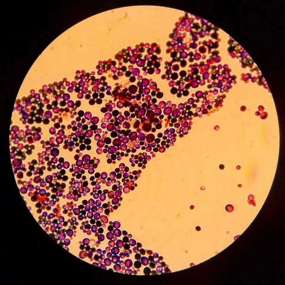
Cancer cells disarm T cells by stealing the molecules they need to fight
Blocking a tumor antioxidant restored T cell activity and made some resistant cancers more responsive to immunotherapy in mice.

 Edited By: Joseph Shavit

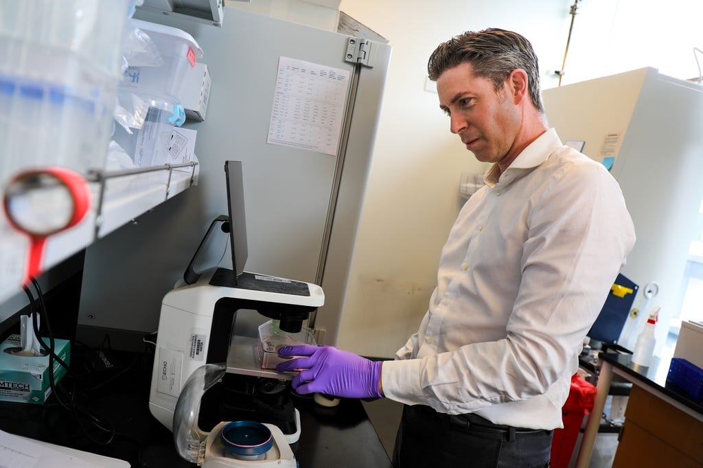
Robert L. Eil, M.D., is co-senior author of a new study in Science that identifies a previously unknown way tumors escape immune attack and points to a promising new target for cancer immunotherapy. (CREDIT: OHSU/Christine Torres Hicks)
- Cancer cells can create an antioxidant-rich environment that deprives tumor-fighting T cells of reactive oxygen species, or ROS, which those immune cells need for normal activation and signaling.
- The antioxidant enzyme PRDX1 emerged as a key suppressor: removing it from cancer cells increased T cell activity, slowed some tumors and made resistant tumors more responsive to checkpoint immunotherapy in mouse models.
- The findings are preclinical and do not establish a new treatment yet, but they identify a previously unrecognized redox checkpoint that could become a target for future cancer immunotherapies.
Reactive oxygen species have spent decades with a bad reputation. Often called free radicals, these chemically reactive molecules can damage DNA, disrupt cells and contribute to cancer when they accumulate in excess.
But cancer-fighting T cells appear to need some of them.
A new study found that tumors can exploit that dependence by flooding their surroundings with antioxidant proteins that remove ROS from the local environment. The result is a chemical barrier that weakens the signaling T cells need to activate, multiply and attack cancer.
The research, published in Science, was co-led by researchers at Oregon Health & Science University and the University of Cambridge.
“One of the surprising findings is that antioxidants aren't always beneficial in the context of cancer,” said Robert L. Eil of the OHSU Knight Cancer Institute. “While reactive oxygen species sound threatening, T cells actually need them to perform their tumor-fighting job.”
Tumors create an antioxidant-rich environment
The researchers began by examining tumor interstitial fluid, the liquid surrounding cells inside tumors.
When fluid collected from mouse melanomas or colorectal tumors was added to activated CD8+ T cells, it strongly suppressed immune functions. The T cells produced less interferon-gamma and tumor necrosis factor and showed reduced signs of cytotoxic activity.
The same effect appeared in fluid from lungs containing melanoma metastases compared with fluid from healthy lungs. Blocking familiar suppressive pathways such as PD-1, CTLA-4 or TGF-beta did not reverse the effect, suggesting another mechanism was involved.
The researchers then measured the fluid's antioxidant capacity and found it was substantially higher than matched serum. T cells exposed to the tumor fluid also showed lower levels of both total and mitochondrial ROS.
That observation suggested the tumor environment was not simply sending a conventional inhibitory signal. It was changing the chemistry around the immune cells.
One antioxidant protein stood out
Protein analysis identified nine known antioxidant proteins in the tumor fluid, but one was especially enriched: peroxiredoxin 1, or PRDX1.
PRDX1 normally helps cells control oxidative stress by neutralizing hydrogen peroxide. Inside cells, that function can be protective. Outside tumor cells, however, it appeared to have a very different consequence.
The researchers estimated PRDX1 concentrations in tumor interstitial fluid at about 0.95 milligrams per milliliter, with most of the protein present in its catalytically active reduced form. Its recycling partners, thioredoxin 1 and thioredoxin reductase 1, were also enriched.
When purified PRDX1 was added directly to T cells, their ROS levels dropped. The cells then produced fewer effector molecules and proliferated less efficiently.
Alexander J. Wesolowski of the University of Cambridge said the findings add to a changing understanding of ROS biology.
“We tend to think of reactive oxygen species purely as damaging byproducts of metabolism,” he said. “But we are increasingly understanding that ROS have important functions within cells, and T cells require them to activate.”
ROS act as part of the T cell’s signaling system
The study helps explain why removing too much ROS can weaken an immune response.
When a T cell recognizes a target, its receptor activates a network of enzymes that relay the signal deeper into the cell. ROS participate in that process by temporarily altering enzymes called phosphatases.
Those phosphatases normally remove phosphate groups from signaling proteins and help shut down activation pathways. Small amounts of ROS can reversibly inhibit them, allowing kinase signaling to remain active long enough for the T cell response to build.
PRDX1 interfered with that balance.
By removing extracellular hydrogen peroxide, the antioxidant protein reduced ROS inside T cells and allowed redox-sensitive phosphatases to remain active. Downstream signaling through pathways involving ERK, AKT and other proteins was then weakened.
The initial triggering of the T cell receptor still occurred. The problem emerged later, when the cell needed to amplify and sustain the signal.
The researchers describe this system as a possible redox checkpoint, with the local balance between oxidants and antioxidants helping determine whether a T cell fully activates.
Removing PRDX1 made some tumors vulnerable
To test whether the mechanism mattered inside tumors, the researchers used CRISPR gene editing to eliminate Prdx1 from several cancer cell lines.
Tumors grown from those edited cells contained much less PRDX1 in their interstitial fluid. Their surrounding fluid also became less effective at suppressing T cells.
The biological effects varied by tumor type.
In one YUMM2.1 melanoma model, PRDX1-deficient tumors grew much more slowly and were rejected by the immune system. Depleting CD4+ and CD8+ T cells reversed that benefit, showing that immune cells were responsible for the tumor control.
In B16-F10 melanoma, removing PRDX1 alone did not substantially change tumor growth. However, these tumors are normally resistant to combined anti-PD-1 and anti-CTLA-4 checkpoint blockade. Once PRDX1 was deleted, they became more responsive to the immunotherapy.
A hepatocellular carcinoma model also showed reduced tumor growth after PRDX1 loss, with a stronger effect when checkpoint blockade was added. A colorectal cancer model, however, did not show the same benefit.
That variation suggests PRDX1 will not be equally important in every cancer.
Evidence also appeared in human cancers
The researchers examined whether the same pattern could exist in people.
Data from 23 human cancer cell lines representing 11 cancer types showed that PRDX1 was one of the most abundant antioxidant proteins released by nearly all of them. Across 21 cancer types in The Cancer Genome Atlas, PRDX1 expression was significantly higher in tumors than adjacent normal tissue in 14.
Tumor fluid collected from two people with melanoma also contained substantially more PRDX1 than matched serum. Human CD8+ T cells exposed to extracellular PRDX1 showed the same suppressive response seen in mouse experiments.
The researchers also found evidence that tumors may increase PRDX1 as they adapt to immune pressure. Cancer cells that had undergone immunoediting expressed more PRDX1 than comparable cells that evolved without the same adaptive immune pressure.
A new target, but not yet a treatment
The study does not show that patients should take pro-oxidant drugs or avoid antioxidants in food. It remains preclinical, and altering ROS throughout the body could produce harmful effects because these molecules can both support signaling and cause cellular damage.
Instead, the researchers are interested in targeting the antioxidant system specifically within tumors. Possible approaches could include blocking extracellular PRDX1, interfering with the thioredoxin system that keeps it active or engineering T cells that resist antioxidant-rich tumor environments.
“What's exciting is that we've identified a target nobody was really looking for before,” Eil said. “This doesn't put a drug in patients' hands tomorrow, but it gives us an entirely new pathway to pursue.”
The findings illustrate why the biology of ROS is more complicated than simply dividing molecules into harmful oxidants and protective antioxidants. Inside a tumor, the same chemistry that protects one cell can weaken another.
For cancer immunotherapy, restoring that balance could eventually give T cells another chance to fight.
Dig deeper into ROS, T cells and cancer immunity
These resources explore how reactive oxygen species influence T cell signaling, exhaustion and the tumor immune environment.
Mitochondria-derived hydrogen peroxide selectively enhances T cell receptor-initiated signal transduction: Shows how hydrogen peroxide produced during T cell activation can act as a signaling molecule that strengthens T cell receptor pathways. (Journal of Biological Chemistry, 2013)
Reactive Oxygen Species: Involvement in T Cell Signaling and Metabolism: Reviews how ROS regulate T cell activation, metabolism and effector functions, providing background for why completely removing these molecules can impair immunity. (Trends in Immunology, 2018)
Glutathione Primes T Cell Metabolism for Inflammation: Demonstrates how antioxidant control and redox balance are tightly linked to the metabolic reprogramming required for effective T cell responses. (Immunity, 2017)
The phosphatase PAC1 acts as a T cell suppressor and attenuates host antitumor immunity: Examines how redox-sensitive signaling and phosphatase activity can shape T cell exhaustion and antitumor immunity inside tumors. (Nature Immunology, 2020)
Mitochondrial stress induced by continuous stimulation under hypoxia rapidly drives T cell exhaustion: Shows that excessive ROS can also become harmful to tumor-infiltrating T cells, highlighting the importance of maintaining the right redox balance rather than simply increasing or decreasing ROS. (Nature Immunology, 2021)
Research findings are available online in the journal Science.
The original story "Cancer cells disarm T cells by stealing the molecules they need to fight" is published in The Brighter Side of News.
Related Stories
- The thymus trains T cells like an AI model to avoid attacking the body
- New injectable system reprograms cancer-fighting T cells inside the body
- 'Superior memory' helps reprogrammed T cells hit tumors harder
Like these kind of feel good stories? Get The Brighter Side of News' newsletter.
Mac Oliveau
Writer
Mac Oliveau is a Los Angeles–based science and technology journalist for The Brighter Side of News, an online publication focused on uplifting, transformative stories from around the globe. Having published articles on MSN, and Yahoo News, Mac covers a broad spectrum of topics including medical breakthroughs, health and green tech. With a talent for making complex science clear and compelling, they connect readers to the advancements shaping a brighter, more hopeful future.