For the first-time ever, scientists image the 3D wavefunction of an organic molecule
A new laboratory method reconstructs electron orbitals at sub-angstrom resolution while sharply reducing measurement demands.
 University of Göttingen Writer: Melissa Sollich

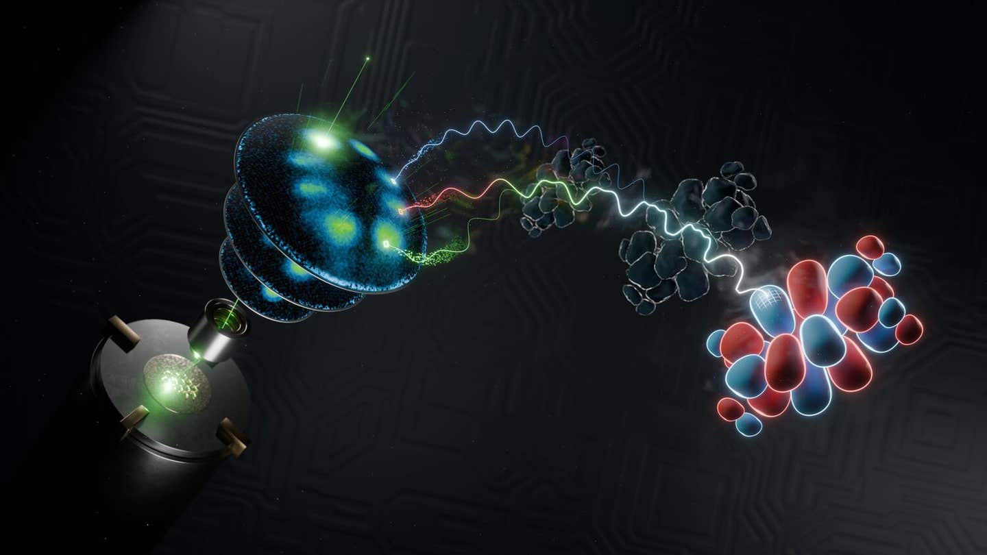
An illustration of how researchers used state-of-the-art photoelectron spectroscopy (left hand side) with a lab-based soft-X-ray light source that provides ultrashort light pulses, which was combined with powerful mathematical algorithms, to image the wavefunction of electron orbitals (right-hand side). (CREDIT: Lukas Kroll)
- Scientists reconstructed a molecule’s full three-dimensional electron wavefunction using a laboratory system instead of a large synchrotron facility.
- A new algorithm produced reliable orbital images from as few as four photon energies, cutting the required measurement time to eight hours.
- The method could eventually record femtosecond-scale changes in molecules, but the current experiment captured only static orbitals.
A molecule’s electron cloud is not a fuzzy picture of where particles sit. It is a three-dimensional quantum map, and scientists have now reconstructed that map in a laboratory with far less data than earlier methods required.
The University of Göttingen team imaged the complete three-dimensional wavefunction of a nanometer-sized organic molecule. The work combined photoelectron spectroscopy with a new reconstruction algorithm and a lab-based source of ultrashort extreme-ultraviolet light.
Wavefunctions sit at the center of quantum mechanics. They describe the possible positions and momenta of particles such as electrons. Inside molecules, these electron wavefunctions form molecular orbitals, which help determine chemical bonding, electronic structure, light absorption, and interactions with nearby materials.
“The wavefunction is a fundamental quantity in quantum mechanics, yet it cannot be directly observed or measured,” said Professor Stefan Mathias of the University of Göttingen.
That problem has forced scientists to work indirectly. In photoelectron spectroscopy, light ejects electrons from a material. Instruments then measure their energy and momentum. Those measurements reveal part of the molecular orbital without directly disturbing the original wavefunction.
Rebuilding what the detector cannot see
The method is called three-dimensional photoemission orbital tomography, or 3D-POT. It records photoelectron patterns at several photon energies. Each measurement captures a curved shell within the molecule’s three-dimensional momentum distribution.
Combining those shells can reveal the orbital’s real-space shape. Earlier versions needed many photon energies and long measurements at synchrotron facilities. That made the technique difficult to use widely and poorly suited for tracking rapid changes.
The Göttingen group reduced both obstacles. Its table-top high-harmonic generation source produced selectable photon energies from 13 to 71 electron volts. The system delivered 35-femtosecond pulses at 500,000 pulses per second.
A time-of-flight momentum microscope recorded the momentum and kinetic energy of each accepted electron. Unlike scanning systems, it collected a broad range of information at once.
The team tested the approach using PTCDA, an organic molecule whose full name is perylene-3,4,9,10-tetracarboxylic dianhydride. The molecules were arranged on a silver surface known as Ag(110).
That interface gave the researchers access to two important orbitals. They imaged the highest occupied molecular orbital, or HOMO, and the lowest unoccupied molecular orbital, or LUMO. Charge transfer from the silver filled the LUMO, allowing both orbitals to appear.
Four photon energies can be enough
The researchers collected data at 10 photon energies between 20.5 and 63.8 electron volts. Each spectrum took about two hours, with a typical rate of 200,000 detected photoelectrons per second.
The central advance came from the reconstruction process. Standard methods depend on closely spaced measurements and interpolation. That approach can create errors near points where the orbital changes sign.
The new algorithm recovered both amplitude and phase, including the orbital’s positive and negative regions. It also estimated parts of momentum space that the experiment did not directly measure.
Dr Matthijs Jansen, a co-leader of the study, said the work joined two advances. “We introduce two powerful new concepts. First, by redesigning the computer algorithm from the ground up, reliable 3D images can now be obtained using much less experimental data. Second, the experiment is based upon a powerful, lab-based soft-X-ray light source that provides ultrashort light pulses. It is the combination of these two techniques that has this remarkable impact.”
Only four photon-energy data sets could produce a complete reconstruction. Those measurements required eight hours. Seven photon energies provided a stronger balance between shorter collection time and reconstruction reliability for the static orbitals.
More measurements improved confidence. With four energies, the algorithm produced more competing solutions. With seven or 10, inaccurate reconstructions were easier to separate from the most likely orbital.
Images sharper than the distance between atoms
The reconstructed orbitals closely matched density functional theory calculations for the isolated molecule. Agreement appeared both across the molecule and in the vertical direction above and below its plane.
The experiment achieved an intrinsic spatial resolution of about 0.75 angstrom. One angstrom equals one ten-billionth of a meter. That resolution is smaller than the spacing between carbon atoms in the molecule.
The method could not resolve every structural detail. Earlier work reported that oxygen atoms in PTCDA sit about 0.3 angstrom closer to the silver than the carbon backbone. That difference falls below the experiment’s resolution.
The researchers found no significant evidence that this slight bending changed the measured HOMO or LUMO. Any effect remained below the detection limit.
The algorithm also depended on constraints, including the molecule’s symmetry, approximate size, and expected orbital sparsity. Releasing some constraints created many possible reconstructions, making the result harder to identify with confidence.
Those limits matter because the larger goal is not only to capture static orbitals. Ultrashort pulses could eventually show how wavefunctions change after optical, electronic, or chemical disturbances.
Practical implications of the research
A laboratory-scale 3D-POT system could make orbital imaging available without relying on a large synchrotron facility. Researchers could examine how organic molecules interact with metals, how electronic states mix at interfaces, and how excitons move through organic semiconductors.
The shorter data requirements also make time-resolved experiments more realistic. The team estimated that four photon energies measured across 10 pump-probe delays would take about 80 hours, comparable with other momentum-microscopy studies.
Dr Wiebke Bennecke, the study’s first author, said the method could move orbital imaging toward atomic-scale movies. “This technique might mean that stroboscopic videography becomes a reality, allowing us to observe not just the shape of wavefunctions, but also to see how it changes with ultrafast, even femtosecond or one quadrillionth of a second, resolution. This will mean we can learn how a molecule adapts to optical, electronic, or chemical changes and find new ways to control these interactions at the level of a few atoms.”
The current work does not yet provide those movies. It establishes the laboratory tools, reconstruction method, and measurement strategy needed to pursue them.
Research findings are available online in the journal Nature Communications.
The original story "For the first-time ever, scientists image the 3D wavefunction of an organic molecule" is published in The Brighter Side of News.
Related Stories
- Organic molecules can survive violent supernova explosions - fueling star and planet formation
- JWST finds a surprise flood of organic molecules in a hidden galaxy
- New AI tool can generate millions of new molecules
Like these kind of feel good stories? Get The Brighter Side of News' newsletter.
Rebecca Shavit
Writer
Based in Los Angeles, Rebecca Shavit is a dedicated science and technology journalist who writes for The Brighter Side of News, an online publication committed to highlighting positive and transformative stories from around the world. Having published articles on MSN, AOL News, and Yahoo News, Rebecca's reporting spans a wide range of topics, from cutting-edge medical breakthroughs to historical discoveries and innovations. With a keen ability to translate complex concepts into engaging and accessible stories, she makes science and innovation relatable to a broad audience.