MIT’s self-organizing laser revolutionizes 3D imaging of the brain’s protective barrier
A chaotic laser beam can self-organize into a sharp pencil beam, giving faster 3D views of the blood-brain barrier.

 Edited By: Joseph Shavit
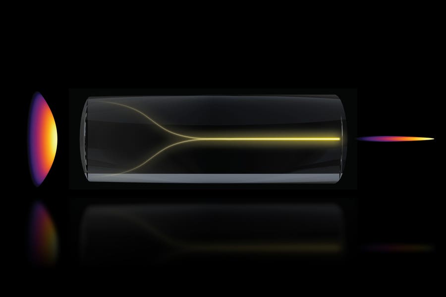
Under the right conditions, a chaotic mess of laser light can spontaneously self-organize into a highly focused “pencil beam.” This schematic shows the pencil beam formation mechanism. (CREDIT: MIT Researchers)
At high power, laser light inside a multimode optical fiber is supposed to misbehave. The beam usually breaks into a noisy, scattered pattern as the light ricochets through many paths at once. But MIT researchers found a case where that expectation fails. Push the system close to its limit, line the beam up just right, and the optical mess can collapse into a tightly focused, self-organized “pencil beam.”
That beam, the team reports, can do more than tidy up a physics problem. In experiments on a human model of the blood-brain barrier, it produced 3D images about 25 times faster than a standard approach. This was achieved while keeping similar cellular-level detail. The work appears in Nature Methods.
“The common belief in the field is that if you crank up the power in this type of laser, the light will inevitably become chaotic. But we proved that this is not the case. We followed the evidence, embraced the uncertainty, and found a way to let the light organize itself into a novel solution for bioimaging,” said Sixian You, an assistant professor in MIT’s Department of Electrical Engineering and Computer Science, a member of the Research Laboratory for Electronics, and the paper’s senior author.
The study was led by MIT graduate student Honghao Cao. Other authors include Li-Yu Yu, Kunzan Liu, Sarah Spitz, Francesca Michela Pramotton, Federico Presutti, Zhengyu Zhang, Subhash Kulkarni of Harvard University and Beth Israel Deaconess Medical Center, and Roger Kamm of MIT.
The beam did the opposite of what it should
The finding began with an observation that seemed wrong.
Cao was testing how much power a multimode fiber could handle. These fibers can carry a great deal of power, but disorder is built into them. Under normal expectations, adding more power should make the output more scrambled, not less.
Instead, when the team drove the laser close to the level that could damage the fiber, the output sharpened into a central peak. The researchers found that this happened only under two strict conditions. The laser had to enter the fiber perfectly on axis, at zero degrees. The power also had to be high enough for the light to interact strongly with the glass itself.
“At this critical power, the nonlinearity can counter the intrinsic disorder, creating a balance that transforms the input beam into a self-organized pencil beam,” Cao said.
The effect emerged in a standard 50-micrometer-core silica step-index multimode fiber. Near the critical power, the light formed a localized beam with a full width at half maximum of 2.5 micrometers, close to the fiber’s diffraction limit of 2.3 micrometers. Its output pulse duration measured 212 femtoseconds, close to a nearly nondispersed reference pulse.
Beam stability
The team also found the beam was unusually stable. Compared with the disordered nonlinear output seen when the beam was launched off axis, the localized state reduced relative power fluctuations by 11 decibels. Under controlled bending tests, it also cut spatial variation 19-fold.
The fiber remained stable over 12 hours without laser-induced damage under the operating condition used for localization. However, the researchers reported irreversible damage when they pushed the power to around 5.5 megawatts.
“That is the charm of this method — you could do this with a normal, optical setup and without much domain expertise,” You said.
Why the cleaner beam matters for imaging
The appeal of the new beam is not only that it forms on its own. It also behaves well in a microscope.
In multiphoton imaging, scientists often face a tradeoff. A tightly focused beam gives good lateral resolution, but only across a thin slice of depth. To build a fuller 3D picture, they have to scan section by section. Existing extended-focus methods, including Bessel-like beams, can cover more depth, but they often bring along sidelobes. These are blurry rings of light that wash out detail and become more sensitive to aberrations.
The MIT beam handled that differently. In point spread function tests with 1-micrometer fluorescent beads, it kept lateral confinement close to the reference laser beam while stretching the axial profile about tenfold. In mouse intestinal tissue labeled with tdTomato, the localized beam produced clear images across an extended axial range in a single frame. For comparison, a conventional Gaussian beam needed a 50-micrometer stack to show similar structure.
The power demands also shifted. Under the same imaging conditions, the speckled multimode output needed 25 milliwatts, the localized beam needed 9 milliwatts, and the laser reference needed 6 milliwatts. However, this was only after 25 repeated z-scans. According to the paper, the localized beam allowed a volume to be imaged with 25-fold shorter acquisition time and with lower photodosage than conventional Gaussian scanning would require.
“Usually, you have a tradeoff between image resolution and depth of focus — you can only probe so far at a time. But with our method, we can overcome this tradeoff by creating a pencil-beam with both high resolution and a large depth of focus,” You said.
A faster look at the blood-brain barrier
The researchers then turned the system toward a harder biological problem: watching transport in a live, intact human blood-brain barrier model.
This barrier protects the brain from toxins, but it also blocks many drugs. In the team’s microfluidic model, human induced pluripotent stem cell-derived endothelial cells, pericytes, and astrocytes were embedded in a fibrin hydrogel matrix. The researchers perfused Alexa555-conjugated transferrin through the microvascular networks. They collected 3D volumes every minute for 50 minutes.
Here the speed mattered. The self-localized beam imaged a 2 millimeter by 2 millimeter by 50 micrometer volume about 25 times faster than a previous setting. This reduced acquisition time from 25 minutes to 1 minute per volume while preserving subcellular resolution.
That let the team see something bulk assays and slower scans miss. Endothelial cells dominated transferrin uptake and reached a plateau around 45 minutes, consistent with classical measurements of transferrin receptor-mediated endocytosis. Pericytes stayed below 30 percent of endothelial uptake, and astrocytes showed little bulk accumulation. But at the single-cell level, behavior varied sharply. Some endothelial cells absorbed transferrin within minutes, while neighboring cells exposed to the same lumen stayed largely quiet.
Transient “hotspots”
The study also reported occasional transient “hotspots” at astrocytic endfeet and found that regions near pericyte coverage often correlated with reduced endothelial uptake. A competitive inhibition assay using excess unlabeled transferrin cut endothelial signal fivefold after 20 minutes. This supported the receptor-mediated nature of the uptake.
“The pharmaceutical industry is especially interested in using human-based models to screen for drugs that effectively cross the barrier, as animal models often fail to predict what happens in humans. That this new method doesn’t require the cells to have a fluorescent tag is a game-changer. For the first time, we can now visualize the time-dependent entry of drugs into the brain and even identify the rate at which specific cell types internalize the drug,” Kamm said.
“Importantly, however, this approach is not limited to the blood-brain barrier but enables time-resolved tracking of diverse compounds and molecular targets across engineered tissue models, providing a powerful tool for biological engineering,” Spitz added.
Practical implications of the research
The work points to a simpler way to get fast, high-resolution volumetric imaging without custom beam-shaping optics or adaptive correction hardware. Because the beam can be generated in a standard step-index multimode fiber, the authors say it could be retrofitted into existing multiphoton microscopes.
For drug research, the immediate value is speed with detail. The method gives scientists a way to watch how cells in a human blood-brain barrier model take up compounds over time, rather than relying only on endpoint snapshots. That could help preclinical screening of brain-targeted biologics, nanoparticles, and gene-delivery systems.
The study also comes with limits. The authors say they still need a fuller theoretical framework to explain the beam’s behavior. They note that certain technical constraints in resolution may contribute to the variability seen in transferrin uptake, and they say visualizing the complete transcytotic pathway across endothelial cells remains difficult because of sensitivity limits. Current imaging speed is also capped by the 1-megahertz laser repetition rate and by the fiber’s energy conversion ratio.
Even so, the paper makes a practical case. A phenomenon that once looked like trouble in high-power fiber optics may turn out to be useful, especially when researchers need to watch biology unfold in three dimensions and in real time.
Research findings are available online in the journal Nature Methods.
The original story "MIT's self-organizing laser revolutionizes 3D imaging of the brain’s protective barrier" is published in The Brighter Side of News.
Related Stories
- Laser-powered, ‘metajets’ could be the future of interstellar flight
- Quantum researchers created a new kind of laser built from sound
- Scientists develop laser-powered graphene propulsion for next-generation space travel
Like these kind of feel good stories? Get The Brighter Side of News' newsletter.
Mac Oliveau
Writer
Mac Oliveau is a Los Angeles–based science and technology journalist for The Brighter Side of News, an online publication focused on uplifting, transformative stories from around the globe. Having published articles on MSN, and Yahoo News, Mac covers a broad spectrum of topics including medical breakthroughs, health and green tech. With a talent for making complex science clear and compelling, they connect readers to the advancements shaping a brighter, more hopeful future.