New microscope captures living cells in stunning 5D detail
New MOSAIC microscope combines 12 imaging technologies and may help create AI systems that transform biological research.

 Edited By: Joseph Shavit
Researchers at UC Berkeley have developed MOSAIC, a powerful microscope that captures life in unprecedented detail and could help build an AI assistant for biology. (CREDIT: Berkeley University)
Deep inside a small, windowless room at the University of California, Berkeley, two microscopes are quietly capturing some of the most detailed views of life ever recorded. Day and night, they collect enormous streams of information, revealing how molecules move, how cells communicate, and how living tissues repair themselves.
The machines, called MOSAIC, may represent a major turning point for biology. More than just microscopes, they are sophisticated imaging platforms that combine a dozen advanced technologies into a single system. Their creators believe they could help launch a new era in biological discovery, one in which artificial intelligence becomes an essential scientific partner.
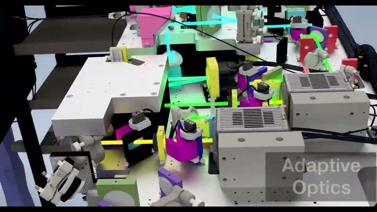
MOSAIC stands for Multimodal Optical Scope with Adaptive Imaging Correction. The system allows researchers to switch between different imaging modes with the press of a button, eliminating the need for multiple specialized microscopes.
Its impact is already spreading. More than a dozen laboratories worldwide have recreated the instrument using detailed assembly instructions shared by the Berkeley team over the past six years.
A New Way to Observe Living Systems
For generations, scientists have relied on different microscopes for different tasks. One instrument might excel at viewing deep tissue. Another might capture fine details at extremely high resolution. Yet each comes with trade-offs.
This creates a challenge for researchers trying to understand living systems across many scales at once.
Life unfolds across vast ranges of size and time. Proteins move in milliseconds. Cells divide over hours. Organisms develop over days. Traditional microscopes often force scientists to focus on only one piece of that picture.
MOSAIC aims to change that.
The system combines multiple imaging methods into a single platform. These include light-sheet microscopy, super-resolution microscopy, multiphoton imaging, label-free imaging, structured illumination microscopy, and adaptive optics.
Together, these tools allow researchers to study everything from individual molecules to entire living organisms.
“Life has to be studied in living tissue, holistically, and over fast timescales and for long periods of time,” said Eric Betzig, a Berkeley professor of molecular and cell biology and physics who received the 2014 Nobel Prize in Chemistry for developing super-resolution fluorescence microscopy.
“You can’t study something as complex as a cell or organism just by looking at the parts individually — there are something like 40 million protein molecules alone of 20,000 different types. With our microscopes, we can image everything from single molecules to whole organisms at high resolution, following as many players as we can to understand natural physiological interactions in the cell.”
Turning Biology Into Five-Dimensional Data
The images produced by MOSAIC are far more complex than ordinary photographs.
Betzig describes the information as five-dimensional. It includes three dimensions of space, along with time and color.
The color comes from fluorescent markers that allow scientists to follow different cellular structures simultaneously. Researchers can track membranes, organelles, protein networks, and other cellular components as they move, change shape, divide, and interact.
The result is an extraordinarily rich view of life in motion.
One experiment followed kidney cells continuously for 24 hours. Researchers collected nearly 1,000 time points and generated roughly 49 terabytes of information. The microscope captured both normal cell divisions and rare events, including abnormal divisions that produced three daughter cells instead of two.
Another experiment observed living zebrafish as they regenerated damaged tail tissue. Researchers watched tiny biological events unfold inside living tissue, including the release of communication vesicles, shifts in structural fibers beneath the skin, fusion between repair cells, and the temporary trapping of a red blood cell as new blood vessels formed.
These observations would have been extremely difficult to capture using older imaging approaches.
Correcting Nature’s Optical Distortions
One of MOSAIC’s most important features is its ability to overcome a longstanding problem in microscopy.
As light travels through living tissue, it becomes distorted. These distortions blur images and reduce detail, especially when researchers look deeper inside organisms.
To solve this problem, MOSAIC uses adaptive optics.
At the heart of the system sits a deformable mirror controlled by 69 tiny motors. These motors continuously adjust the mirror’s shape to compensate for distortions caused by tissue.
The corrections sharpen images in real time, allowing researchers to maintain clarity even deep inside living specimens.
The microscope can image neural structures inside live mouse brains, follow cancer cells moving through zebrafish blood vessels, and map nanoscale structures across expanded human brain tissue.
In one study involving mouse brains, adaptive optics helped researchers detect more than twice as many calcium signaling events compared with imaging without correction.
The Growing Data Challenge
While MOSAIC solves many imaging problems, it creates a new one: data overload.
The system can generate as much as four terabytes of information every hour. A single experiment may produce between 30 and 100 terabytes of data.
Some datasets are measured in petabytes. One petabyte equals roughly 500 billion pages of text.
That volume of information exceeds what scientists can realistically analyze on their own.
“We are the world’s best at collecting data at 5D, and have been for a decade,” Betzig said. “But we don’t know how to interpret the data at scale; we can’t think in petabytes and we don’t see in 5D. That’s why we’re developing a 5D AI — it’s a sherpa to guide us.”
Researchers believe artificial intelligence will become essential for making sense of these massive biological datasets.
Building a ChatGPT for Biology
The Berkeley team is now working toward what they call a Cell Observatory Initiative. The goal is to create an AI system capable of understanding complex biological movies much the way language models understand text.
Srigokul “Gokul” Upadhyayula, who led MOSAIC’s development, believes biology has entered a new phase.
“Biology is entering an era in which the data are too complex and too large to interpret by human inspection alone,” he said. “A biologist may understand the biological question deeply, but still lack the computational tools and infrastructure needed to process, analyze and quantify what they are seeing. We need to build a mind that can reason natively with 3D movies of living biological systems and let us query those dynamics through language — akin to a ChatGPT for biology.”
Such a system could help researchers ask questions directly.
Instead of manually examining thousands of images, scientists might ask how many immune cells entered a wound, when a cell began migrating, or whether certain behaviors predict disease progression.
“There’s so much information in these large movies, across scales, about how cells are behaving in the organism and the tissue and at the subcellular level, it can be difficult even for a very well-trained biologist to understand or digest,” said Ian Swinburne, a Berkeley assistant professor of molecular and cell biology.
“AI can help us interface with the data and ask or answer questions more easily.”
Discovering the Unexpected
One of the most exciting aspects of MOSAIC may be its ability to reveal phenomena researchers were not even looking for.
Because the images contain so much information, scientists frequently notice unexpected biological events while studying something else.
“There’s so much information in these movies,” Swinburne said. “We come in with maybe a hypothesis about the process we think we’re studying and then we get distracted by something we’ve never seen before. Probably every movie has something new that we acquire just because the quality is so high, the spatial and time resolution so much better than what we’re used to.”
For researchers, that unpredictability is part of the appeal.
Many major discoveries begin with observations nobody expected to make.
Practical Implications of the Research
MOSAIC could fundamentally change how scientists study living systems. By combining many imaging technologies into one platform, researchers can examine biological processes across multiple scales without moving samples between different instruments. This creates a more complete picture of how cells, tissues, and organisms function in real time.
The technology may accelerate discoveries in areas such as cancer biology, neuroscience, regenerative medicine, developmental biology, and infectious disease research. Scientists could better understand how tumors spread, how brain cells communicate, how wounds heal, and how diseases alter cellular behavior.
The project may also help drive the development of powerful new AI systems designed specifically for science. These tools could dramatically reduce the time required to analyze complex datasets and help researchers identify patterns that would otherwise remain hidden.
By pairing advanced imaging with intelligent analysis, scientists hope to uncover biological insights that could ultimately improve human health and deepen our understanding of life itself.
Research findings are available online in the journal Nature Methods.
The original story "New microscope captures living cells in stunning 5D detail" is published in The Brighter Side of News.
Related Stories
- A new type of microscope lets scientists observe life unfolding inside cells
- AI microscope autonomously performs tasks like a scientist, only much faster
- SMART technology converts existing mobile phone cameras into high-resolution microscopes
Like these kind of feel good stories? Get The Brighter Side of News' newsletter.
Mac Oliveau
Writer
Mac Oliveau is a Los Angeles–based science and technology journalist for The Brighter Side of News, an online publication focused on uplifting, transformative stories from around the globe. Having published articles on MSN, and Yahoo News, Mac covers a broad spectrum of topics including medical breakthroughs, health and green tech. With a talent for making complex science clear and compelling, they connect readers to the advancements shaping a brighter, more hopeful future.