
Researchers discover a hidden world of living cells in human breast milk
Living cells in human milk handle nutrients in different ways, nanoscale imaging reveals, offering a closer look at milk production and immune cell biology.
 UWA Writer: Annelies Gartner

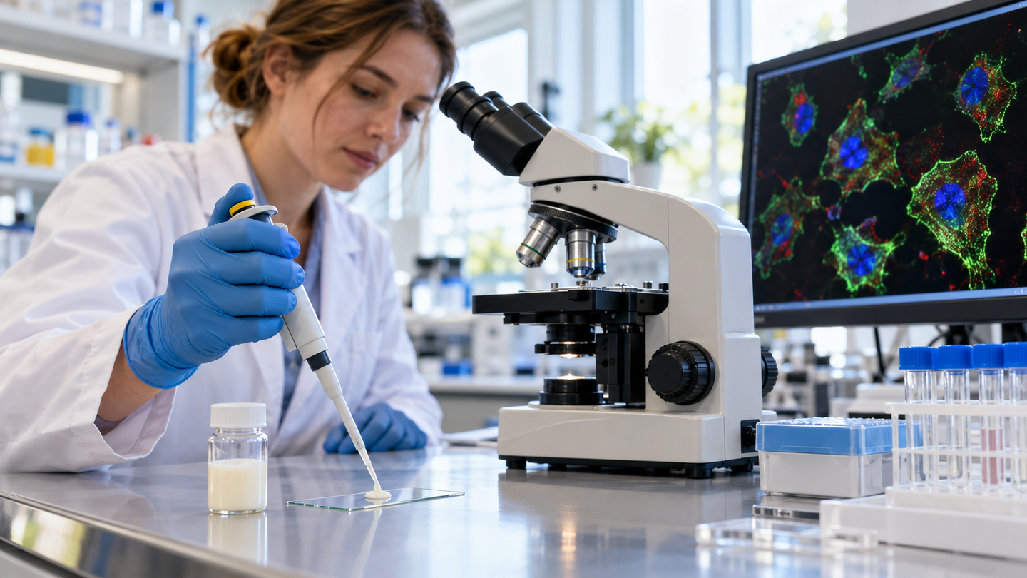
Nanoscale imaging reveals how living cells in human milk differ in structure, chemistry and nutrient handling. (CREDIT: Shutterstock)
- Nanoscale imaging revealed distinct structures and nutrient-handling patterns among living cells in human milk.
- Glucose tracing highlighted lipid-handling immune cells, while amino acid tracing identified differences among milk-producing cells.
- The small laboratory study establishes a research platform, but does not demonstrate effects on infant health.
Living cells in human milk do more than share the same fluid. Some immune cells concentrate carbon from sugar inside fat droplets, while certain milk-producing cells accumulate more material from protein building blocks. A new imaging study reveals these differences in unusually fine detail, giving scientists a closer view of the cellular activity within milk.
The research, co-authored by biomedical scientists at The University of Western Australia and published in ACS Nano, combines several imaging methods with nutrient tracing. Its contribution goes beyond identifying which cells are present. The approach connects cell identity with internal structures and chemical maps, helping reveal how different cells handle nutrients under laboratory conditions.
Human milk was already known to contain living cells, including milk-producing lactocytes and several immune cell types. What remained difficult was seeing their internal organization alongside their metabolic activity, with enough spatial detail to locate nutrient-derived signals inside individual cellular compartments. Milk's abundant fat and varied contents complicate that task, obscuring details that conventional cell profiling cannot fully capture.
Looking inside the same cell
Lead author Qiongxiang, known as Lynn, Lin is a doctoral candidate who studies this cellular complexity. “With our new approach, we can look inside individual cells at the nanoscale and begin to understand how their structure, chemical composition and metabolism are connected,” she said. Those connections matter because cells with similar identities may perform differently.
The team combined confocal microscopy, electron microscopy and nanoscale secondary ion mass spectrometry, or NanoSIMS. Confocal imaging uses fluorescent markers to help identify cell types, while electron microscopy reveals much smaller internal structures. NanoSIMS maps elements and isotopes, which are forms of an element with different numbers of neutrons.
Each technique answers a different question about a cell. By locating the same cells across images, the researchers could connect their identities with features such as fat droplets and chemical concentrations. Gridded surfaces helped align the measurements, reducing the uncertainty that would arise from comparing unrelated cells viewed through different instruments.
The resulting structural survey included lactocytes, macrophages, neutrophils and dendritic cells. The latter three belong to the immune system, although their roles and internal organization differ. Structural analyses included samples from five participants, while the nutrient-tracing measurements relied on two donors, an important limit on how broadly the findings can be applied.
Following sugar into immune cells
To investigate metabolism, the team added glucose containing a stable carbon isotope to freshly collected milk samples. This label allowed them to track where carbon originating in the sugar accumulated. After 30 minutes, NanoSIMS detected markedly different labeling patterns among macrophages, immune cells that can engulf material and help coordinate immune responses.
Some macrophages had much stronger carbon labeling inside their lipid droplets than others. These droplets are cellular compartments that store fats, making the pattern consistent with active lipid handling. The result shows that milk macrophages are metabolically varied, rather than a uniform population doing the same work at the same rate.
However, the images do not establish exactly how the labeled carbon reached those droplets. NanoSIMS identifies isotope enrichment, not the specific molecules carrying it. Additional biochemical measurements would be needed to distinguish newly synthesized fat from lipid remodeling or other pathways, so the findings cannot simply be read as proof that sugar became new fat.
Lactocyte fat droplets showed little glucose-derived labeling during the same experiment. That contrast suggests different short-term nutrient-handling patterns between these cell populations. It does not mean lactocytes never use glucose, because the experiment captured only a particular tracing period and set of laboratory conditions outside the body.
Milk-producing cells have different activity levels
A second experiment followed amino acids, the building blocks of proteins, using a stable nitrogen isotope. The researchers examined labeling over intervals extending from zero to 30 minutes. Individual lactocytes accumulated different amounts of the label, revealing variation even within the cell population associated with producing milk components.
That variation offers a more detailed picture than treating all lactocytes as equally active. Some cells showed substantially greater accumulation of amino-acid-derived nitrogen than others. Yet the measurement cannot distinguish nitrogen incorporated into newly made proteins from free amino acids or other nitrogen-containing molecules within the cells.
The scientists also reanalyzed a publicly available single-cell RNA sequencing dataset to explore gene activity. That technique measures RNA messages associated with genes being expressed. Lactocyte groups with stronger amino acid metabolism signatures also showed patterns associated with protein synthesis, secretion, energy production and lipid metabolism, consistent with their established secretory role.
Crucially, those RNA measurements came from a separate dataset, rather than the exact cells imaged during nutrient tracing. The two approaches therefore provide complementary evidence, not a direct molecular record of every photographed cell. Together, they support metabolic diversity among lactocytes without establishing precisely how much milk protein each imaged cell produced.
Chemical clues to immune defenses
Neutrophils offered another example of how internal structure and chemistry can be studied together. NanoSIMS detected sulfur and iron enrichment within their intracellular granules, small compartments that can carry antimicrobial material. Those elemental signatures fit with known features of neutrophil defenses, although they do not identify the specific proteins involved.
Professor Donna Geddes, director of UWA's Centre for Human Lactation Research and Translation, highlighted the relevance of these immune cells to infection protection. The study provides a way to examine their contents more closely. It did not directly test whether the measured granules killed bacteria or whether their composition changed infection rates in babies.
Sample preparation also proved consequential for interpreting cellular biology. Lactocytes attached to coverslips showed compromised internal structures even when familiar cell markers remained detectable. The team conducted metabolic tracing with freshly isolated cells in suspension to reduce effects associated with attachment and culture, underscoring how handling can influence what an experiment reveals.
Questions about lactation and maternal health
For Dr. Kai Chen of UWA's School of Biomedical Sciences, combining these methods provides a way to connect cell structure with function. The platform could help scientists investigate differences that remain hidden when cells are counted or classified alone. Larger studies will need to determine how consistently these metabolic patterns appear across individuals and throughout lactation.
Associate Professor K. Swaminathan Iyer, director of the ARC Training Centre for Biomedical Analysis, identified gestational diabetes as a future research interest. Whether that condition alters milk cells' metabolism remains unanswered by this study. Testing it would require appropriately designed comparisons, rather than assuming the current findings already reveal differences between mothers with and without diabetes.
The immediate advance is a method for asking more precise questions about living milk cells. It does not establish a clinical test or demonstrate that changing those cells improves infant outcomes. A next step is to identify the labeled molecules themselves and determine whether the same cellular patterns persist across larger, more varied groups of donors.
Dig deeper into living cells in human milk
These studies place the imaging findings within research on milk production, cellular diversity and immune responses.
Genomic characterization of normal and aberrant human milk production
This study connects milk production differences with gene expression and cell composition. It also provides the public single-cell dataset reanalyzed in the imaging research. (Science Advances, September 2025)
Lactation-associated macrophages exist in murine mammary tissue and human milk
Research in mice, with comparisons to human milk cells, explores specialized macrophages associated with lactation. It adds context for interpreting lipid-handling immune cells without equating mouse findings with infant outcomes. (Nature Immunology, June 2023)
Cellular and transcriptional diversity over the course of human lactation
Single-cell sequencing tracks how milk cell populations and gene activity vary across lactation, providing a longer-term perspective than brief nutrient-tracing experiments. (Proceedings of the National Academy of Sciences, April 2022)
Research findings are available online in the journal ACS Nano.
The original story "Researchers discover a hidden world of living cells in human breast milk" is published in The Brighter Side of News.
Related Stories
- Scientists create smart lactation pad to monitor breast milk in real time
- Groundbreaking study links human breast milk to brain health and development
- Antibodies in breast milk help shape infants’ immunity
Like these kind of feel good stories? Get The Brighter Side of News' newsletter.
Joseph Shavit
Writer, Editor-At-Large and Publisher
Joseph Shavit, based in Los Angeles, is a seasoned science journalist, editor and co-founder of The Brighter Side of News, where he transforms complex discoveries into clear, engaging stories for general readers. With vast experience at major media companies like The Los Angeles Times, Times Mirror and Tribune Publishing, he writes with both authority and curiosity. His writing focuses on space science, planetary science, quantum mechanics, geology. Known for linking breakthroughs to real-world markets, he highlights how research transitions into products and industries that shape daily life.