Stanford researchers discover a language-specific network hidden in the human brain
Nearly 2,000 brain scans reveal a stable language system that remains detectable even when people are not reading, speaking, or listening.
 Stanford University Writer: Sara Zaske

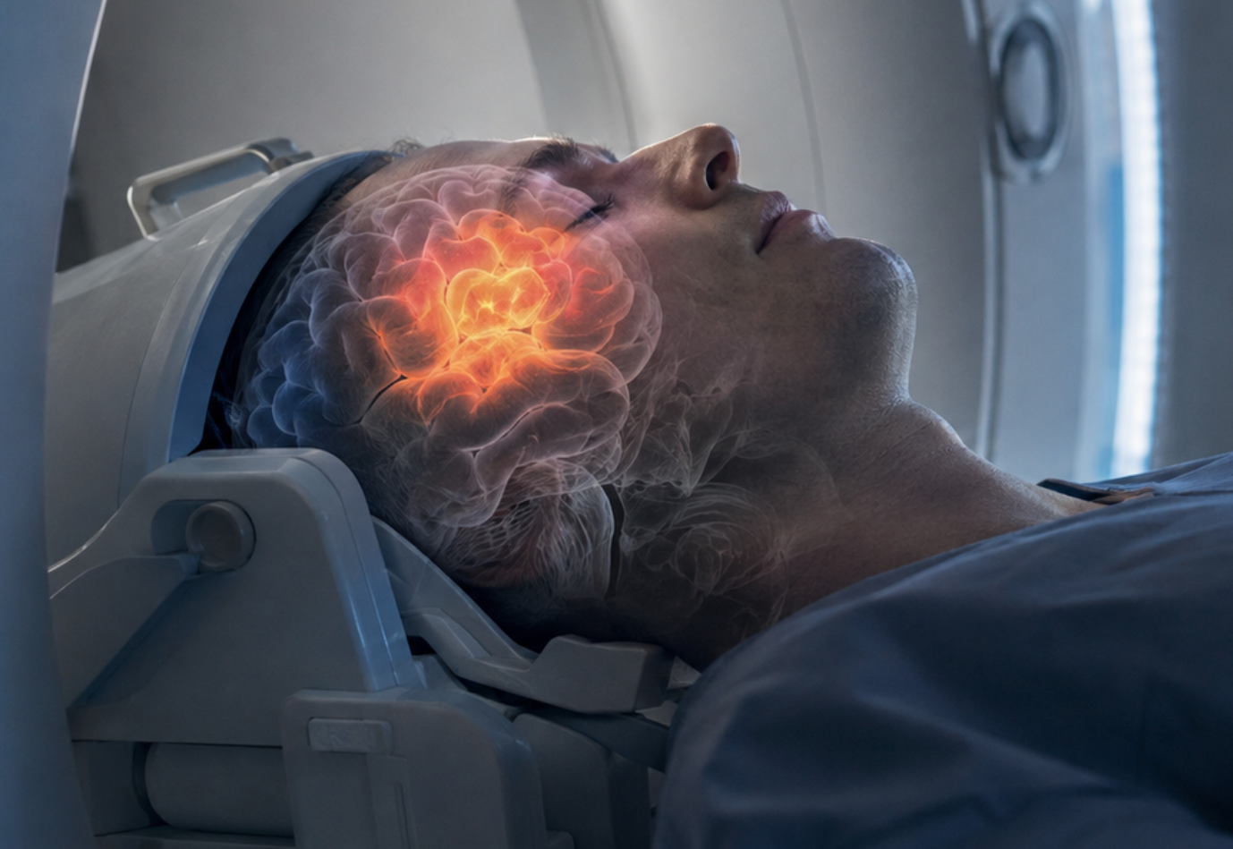
The brain language network can be mapped from fMRI activity even while people rest or perform tasks unrelated to language. (CREDIT: Shutterstock)
- A language-specific brain network can be identified even when a person is resting, listening to music, or doing a task unrelated to language.
- The network is usually concentrated in left frontal and temporal brain regions, but its exact shape varies substantially from person to person.
- The approach could help scientists study existing brain scans and may eventually aid work involving aphasia, neurosurgery, speech prostheses, and brain stimulation.
The brain’s language system does not switch off when conversation stops. Its activity remains organized enough for scientists to identify the network even when someone is solving a visual puzzle, listening to music, or doing nothing in particular.
An analysis of 1,957 functional magnetic resonance imaging sessions from 1,199 people found strong evidence for a distinct network involved in language. The work suggests that the network can be mapped from patterns of brain activity alone.
“The human brain appears to have a network that’s selective and necessary for language, and we can find that network from the activity in the brain alone,” said Cory Shain, lead study author and assistant professor of linguistics in the Stanford School of Humanities and Sciences. “The simple fact that a person’s brain activity is always going up and down lets us detect where this network is with high fidelity. It’s such an important and stable structure that we can find it no matter what someone is doing.”
Shain and co-author Evelina Fedorenko of the Massachusetts Institute of Technology found that the network generally covers frontal and temporal areas, with a strong left-hemisphere bias. Its precise layout, however, differs from one person to another.
A network that survives changing tasks
Language has occupied a central place in brain science since the discovery of aphasia, which can selectively impair speaking or writing after brain injury. Yet scientists still disagree about exactly which areas support language and how those areas divide different linguistic functions.
“Language is a very complex thing,” Shain said. “It's like a spider web that ties together almost everything about us. It’s hard to reach agreement on a definition of language, or even whether language is a unified ‘thing’ at all. With this study, we tried to set aside our preconceptions about what language is and ask what the brain itself is telling us about how language might be implemented.”
The team turned to individualized functional connectomics, which tracks how activity in different brain regions rises and falls together. Functional MRI, or fMRI, measures changes related to blood flow. Because blood flow responds to neural activity, researchers can examine which brain regions fluctuate together over time.
The dataset included every eligible fMRI session collected in Fedorenko’s laboratory from 2007 through 2024. Researchers excluded studies involving people with brain lesions or an established neurodevelopmental diagnosis such as autism.
Rather than begin by asking participants to perform a specific language task, the analysis searched their brain activity for groups of regions showing strongly coordinated patterns.
Language emerges without language stimulation
Participants had completed a variety of activities while being scanned. Some involved language, including reading or listening. Others examined working memory, numerical thinking, music, visual perception, or the ability to reason about other people’s mental states.
The researchers first analyzed changes in brain activity without using information about what each participant was doing. Their automated method repeatedly divided each brain into functionally connected networks and matched those networks with reference patterns.
One network consistently emerged in areas previously associated with language. It responded strongly during language tasks but not during most tasks involving other forms of thought.
The team then removed all scans from tasks involving language input or responses. Even with only nonlinguistic activity, the method recovered a similar network.
Its structure remained stable within the same person across different scanning sessions. At the same time, its shape varied between individuals, strengthening evidence that language regions cannot always be mapped accurately using the same anatomical coordinates for everyone.
Left-sided, selective and tightly connected
The network showed a pronounced left-hemisphere preference. Its laterality index averaged 0.61, while a standard reading-based language localizer produced a nearly identical average of 0.60.
It also behaved differently from neighboring systems.
An auditory network responded strongly during listening. A frontoparietal network responded to working-memory and mathematics tasks while showing weak or negative responses to language. Another nearby network responded most strongly when participants considered other people’s thoughts.
Those differences provided evidence that the language system was not simply part of a broader network handling difficult tasks, sound, or social reasoning.
The language network also remained remarkably intact when the researchers changed how finely they divided the brain. Once the analysis reached roughly 50 networks, its size and shape stabilized. It stayed largely intact even when the brain was divided into as many as 200 networks.
That result supports the idea that the language network operates as a tightly connected functional unit rather than separating neatly into large anatomical regions responsible for different language functions.
One scan can reveal the pattern
More data improved the accuracy of the method, but large amounts were not always necessary.
Among 53 participants with at least 50 imaging runs, the researchers compared network estimates based on different amounts of data. Estimates became more stable as additional runs were included, especially after five or more.
Still, the language network showed selective responses even when researchers estimated it from a single functional run, averaging 198 time points. Additional data strengthened the network’s functional selectivity.
The researchers also tested whether the method could replace a conventional language-localizer scan in an earlier reading experiment involving 40 participants.
Using the connectivity-based network largely reproduced the sensitivity of task-based localization and performed better than simply averaging activity across broad anatomical regions. Task-based localization remained somewhat stronger for one measured effect, but individualized connectivity provided a substantial improvement over group-level anatomical mapping.
Individual differences complicate a universal brain map
The findings challenge approaches that assign language functions to the same large anatomical regions in every brain.
Both the connectivity-derived network and conventional language-task maps remained more similar across repeated scans of the same person than across different people. That pattern suggests each brain has a relatively stable language-network layout, but population averages can blur meaningful differences.
The authors caution that the automated method can still introduce errors. The clustering procedure can settle on imperfect solutions, and the system could occasionally label the wrong network as the language network.
They also tested only one basic approach to dividing functional connectivity patterns into networks. More sophisticated methods could improve localization.
The researchers do not argue that language-localizer tasks should disappear. Such tasks can independently confirm that a mapped network truly responds to language and measure how strongly it responds. When those scans are practical, the two approaches can provide complementary evidence.
Practical implications of the research
Task-independent mapping could make existing fMRI collections more useful because researchers may be able to identify language networks even when older datasets contain no dedicated language-localizer scan. The method may also help when task information is incomplete or when participants cannot tolerate long scanning sessions.
Shain said the approach could eventually contribute to research involving people with brain injuries.
“If we can reliably determine how the damaged areas were organized on the basis of the organization of the rest of the brain that's still intact, then we could discover new things about the causes and consequences of aphasia, which could have clinical relevance for a lot of people,” he said.
The authors also identify possible uses in mapping language-sensitive cortex before neurosurgery, choosing sensor locations for neural speech prostheses, and selecting targets for language-related brain stimulation.
For now, the central result is more fundamental. A person does not need to speak, read, or even think about language for the brain’s language network to leave a recognizable signature in its ongoing activity.
Dig deeper into the brain’s language network
These resources explore how the brain organizes language, why language regions vary between people, and how individualized brain mapping could improve research and clinical applications.
Precision fMRI reveals that the language network exhibits adult-like left-hemispheric lateralization by 4 years of age
Using precision fMRI, researchers found that the specialized frontal and temporal language network already shows strong left-hemisphere lateralization in young children, adding developmental context to the network seen in adults. (Nature Communications, 2026)
A 3.5-minute-long reading-based fMRI localizer for the language network
This work evaluates a shortened fMRI procedure for locating language-responsive regions within individual brains, addressing the practical challenge of obtaining reliable language maps without consuming large amounts of scanning time. (Imaging Neuroscience, 2026)
The language network as a natural kind within the broader landscape of the human brain
This comprehensive review describes a strongly interconnected, largely left-hemisphere frontal and temporal network that is highly selective for language and distinct from systems supporting perception, movement, reasoning and other cognitive functions. (Nature Reviews Neuroscience, 2024)
The Language Network Reliably “Tracks” Naturalistic Meaningful Nonverbal Stimuli
Researchers found that language-network activity can track meaningful nonverbal events, although responses remain weaker than those produced by linguistic material, helping clarify the boundaries of the network’s functional selectivity. (Neurobiology of Language, 2024)
Probabilistic atlas for the language network based on precision fMRI data from >800 individuals
Using individual language-localizer scans from 806 people, researchers created a probabilistic atlas showing substantial person-to-person variation in language-region locations and demonstrating why individualized functional mapping can outperform simple anatomical assumptions. (Scientific Data, 2022)
Research findings are available online in the journal Nature Communications.
The original story "Stanford researchers discover a language-specific network hidden in the human brain" is published in The Brighter Side of News.
Related Stories
- MIT study finds hidden language network in the human brain
- Study finds universal rules behind how languages reuse words
- AI tool builds entirely new languages, then checks its own grammar
Like these kind of feel good stories? Get The Brighter Side of News' newsletter.
Joseph Shavit
Writer, Editor-At-Large and Publisher
Joseph Shavit, based in Los Angeles, is a seasoned science journalist, editor and co-founder of The Brighter Side of News, where he transforms complex discoveries into clear, engaging stories for general readers. With vast experience at major media companies like The Los Angeles Times, Times Mirror and Tribune Publishing, he writes with both authority and curiosity. His writing focuses on space science, planetary science, quantum mechanics, geology. Known for linking breakthroughs to real-world markets, he highlights how research transitions into products and industries that shape daily life.