AI model helps MRI scans predict diabetes, heart risks and death
AI-built MRI body maps show muscle quality, not just fat or BMI, can signal future diabetes, heart events and death

 Edited By: Joseph Shavit
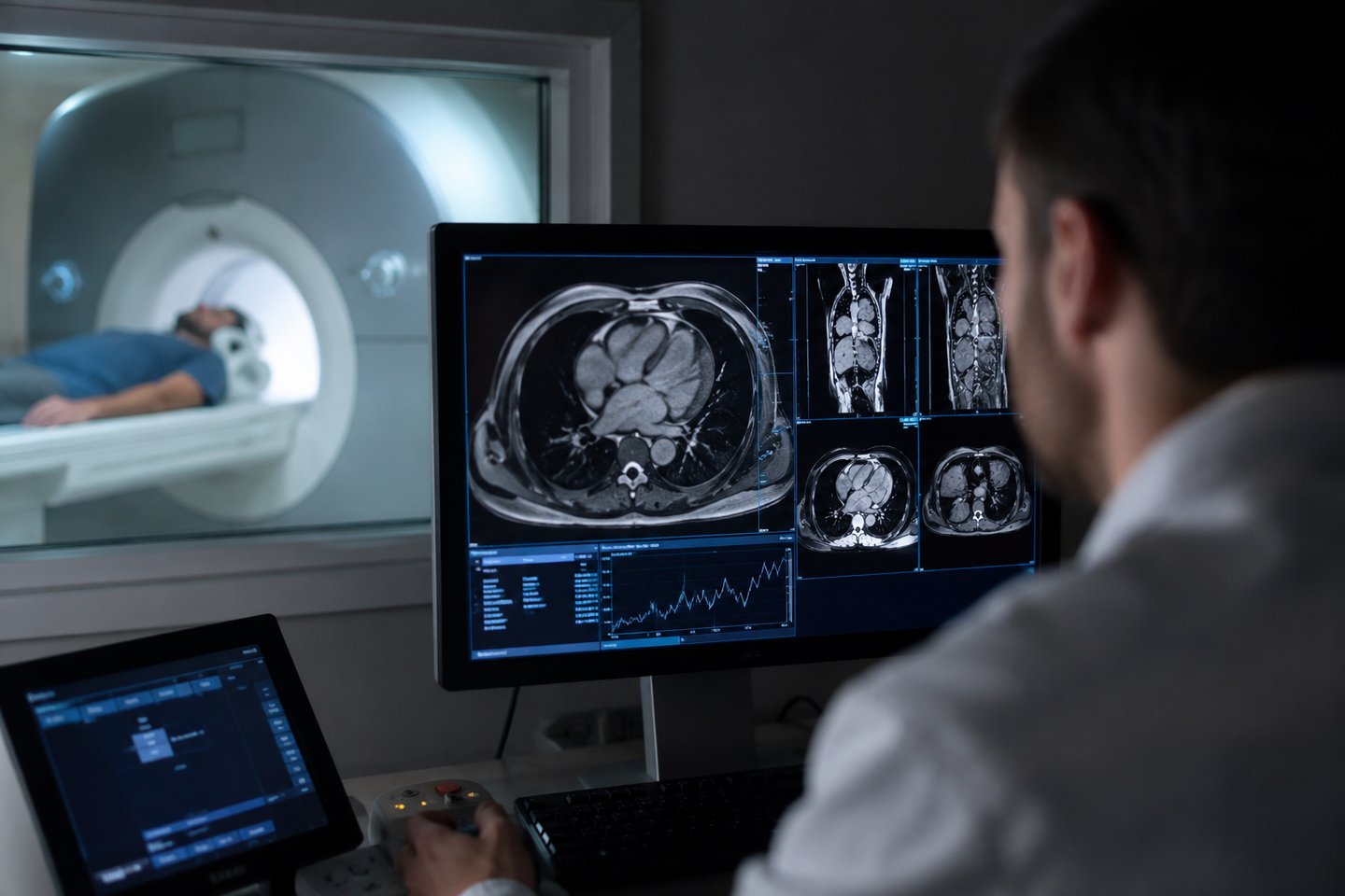
AI analysis of 66,000 MRI scans found muscle quality and visceral fat predict diabetes, heart risk and mortality. (CREDIT: AI-generated image / The Brighter Side of News)
A bathroom scale can tell you how much you weigh. A body mass index chart can sort you into broad categories. However, neither one can show where fat is building up or how much muscle you have left. Nor can they show whether that muscle is quietly filling with fat as you age.
That gap sits at the center of a new imaging study that used artificial intelligence to examine whole-body MRI scans from 66,608 people. The work, published in Radiology, produced a detailed reference map of how fat and muscle are distributed across the body by age, sex and height. Furthermore, it points to something doctors often miss. Muscle quality may matter nearly as much as fat quantity when it comes to predicting disease and death.
The research drew on participants from the UK Biobank and the German National Cohort, two large population studies that gave investigators access to scans collected between April 2014 and May 2022. The average participant was 57.7 years old, and the average BMI was 26.2.
For years, doctors have leaned on BMI and waist size because they are easy to measure. Jakob Weiss, a radiologist at University Medical Center Freiburg in Germany and the study’s senior author, said that convenience comes with a cost.
“Many risk scores and treatment decisions still rely on BMI or waist circumference because they are simple to obtain,” Weiss said. “But BMI does not reliably reflect a person’s actual body composition.”
A body changes in ways BMI cannot track
Using an open-source deep learning system, the team measured several parts of body composition from MRI scans: subcutaneous fat under the skin, visceral fat around the organs, skeletal muscle, skeletal muscle fat fraction, and intramuscular adipose tissue, a marker of fat within muscle.
The researchers then converted those measurements into z-scores, showing how far a person’s body composition differed from the expected range for someone of the same age, sex and height.
That adjustment mattered. Matthias Jung, the study’s first author, said these measures are shaped by normal biology as much as by disease risk.
“There is growing evidence that body composition measures are independent risk factors for cardiometabolic and oncological diseases and mortality,” Jung said. “However, these measures are influenced by height and sex and change substantially with age.”
Across adulthood, the scans showed a steady reshaping of the body. In women, subcutaneous fat increased by 0.42 liters per decade, visceral fat by 0.39 liters, skeletal muscle fat fraction by 0.90 percentage points, and intramuscular fat by 0.19 deciliters. Skeletal muscle, meanwhile, fell by 0.48 liters per decade.
Men followed a similar pattern, though some changes were steeper. Subcutaneous fat rose by 0.30 liters per decade, visceral fat by 0.60 liters, skeletal muscle fat fraction by 0.82 percentage points, and intramuscular fat by 0.17 deciliters. Skeletal muscle dropped by 0.78 liters per decade.
Muscle decline started earlier than many might expect. The analysis found that skeletal muscle began to fall at about age 30 in both sexes. At the same time, muscle fat fraction and intramuscular fat climbed across the lifespan.
Not all fat carries the same warning
The study did more than chart age-related shifts. It also tested which body composition patterns were linked to later illness in the UK Biobank group. In this group, follow-up outcome data were available.
After excluding people who already had diabetes, insulin use, heart attack or stroke before their MRI scan, the outcome analysis included 34,445 individuals. Over a median follow-up of 4.2 years, 532 people developed diabetes, 553 experienced a major adverse cardiovascular event, and 563 died.
The strongest warning sign for future diabetes was high visceral fat. People whose visceral fat was more than one standard deviation above the age-, sex-, and height-adjusted norm had a 2.26-fold higher risk of developing diabetes. This was true even after accounting for age, sex, BMI category, race, smoking, alcohol use, hypertension, blood pressure medication and cancer history.
Muscle quality stood out in a different way. High intramuscular fat was tied to a 1.54-fold increased risk of major cardiovascular events. Low skeletal muscle predicted a 1.44-fold higher risk of death from any cause. High skeletal muscle fat fraction also tracked with higher risks of diabetes and mortality.
That helps explain why two people with similar weight or BMI can face very different futures.
“It’s not only how much muscle you have, but also it’s the quality of that muscle,” Jung said. “Knowing the volume of intramuscular fat gives us a window into muscle quality that other methods like BMI, bioelectrical impedance analysis, or DEXA can’t easily provide.”
The imaging also showed where tissue shifts over time. In both sexes, subcutaneous fat moved from the gluteal region toward the chest with age, while visceral fat shifted from the pelvis toward the abdomen. Paraspinal intramuscular fat moved from the lower lumbar spine toward the upper thoracic spine.
A reference chart for what “normal” really means
One of the study’s biggest contributions may be the reference curves themselves.
Rather than using one cutoff for everyone, the Freiburg team built age-, sex-, and height-adjusted curves for whole-body scans and for more routine scan regions such as the chest, abdomen, pelvis and a single slice at the L3 vertebra. Additionally, they released a public web-based calculator. As a result, researchers and clinicians can compare their own patients or datasets against those benchmarks.
“Adjusting for confounding factors is critical for improving screening accuracy and tailoring treatment decisions,” Weiss said. “This tool has the potential to identify whether an individual’s body composition puts them at greater risk for metabolic disease compared to their age-matched peers.”
That could open the door to a more opportunistic use of scans that already exist. A patient may not need a dedicated whole-body MRI just to assess body composition.
“This tool can allow clinicians to use routine imaging opportunistically,” Weiss said. “A dedicated whole-body MRI is not necessarily required. If a routine CT or MRI body scan already exists, the information can be extracted for benchmarking against the reference values.”
He added that hospitals already capture these signals but rarely measure them.
“We’re already imaging patients every day,” Weiss said. “On every scan of the abdomen or chest, the information is there, we just don’t routinely measure or report it. AI now allows us to tap into this hidden layer of data in a quantitative, reproducible way.”
The study has limits. The cohort was made up mostly of White Western European adults from Germany and the United Kingdom, most older than 20 and slightly overweight. Therefore, the reference curves may not transfer cleanly to other racial or ethnic groups, children, people with very different BMI patterns, or patients in clinical populations. Outcome data were also available only in the UK Biobank.
Practical implications of the research
This work suggests that future risk assessment may move beyond body weight and BMI toward something more specific: where fat sits, how much muscle remains, and how healthy that muscle is.
In practice, that could sharpen screening for diabetes and cardiovascular disease, especially in people whose BMI does not look alarming. It may also matter in cancer care, where body composition can affect treatment tolerance, toxicity, survival and recurrence. Weiss said the approach could even help doctors distinguish healthy fat loss from harmful muscle loss in people using weight-loss drugs such as GLP-1 agonists.
The next step is to test these reference curves in real clinical populations and build disease-specific benchmarks. If that happens, routine scans of the chest or abdomen may start offering more than a look at organs or tumors. They may also provide a running account of how the body itself is changing, and whether those changes are starting to carry a cost.
Research findings are available online in the journal Radiology.
The original story "AI model helps MRI scans predict diabetes, heart risks and death" is published in The Brighter Side of News.
Related Stories
- Brain MRIs and AI predict brain age, cancer survival, and other diseases
- AI tool tracks facial aging, and helps doctors gauge cancer risk
- AI tool predicts brain tumors before surgery with near-perfect precision
Like these kind of feel good stories? Get The Brighter Side of News' newsletter.
Shy Cohen
Writer