FAU scientists discover that shark spines are not one-size-fits-all
Micro-CT scans uncovered how six shark species carry spinal designs tuned to their distinct swimming styles.

 Edited By: Joseph Shavit

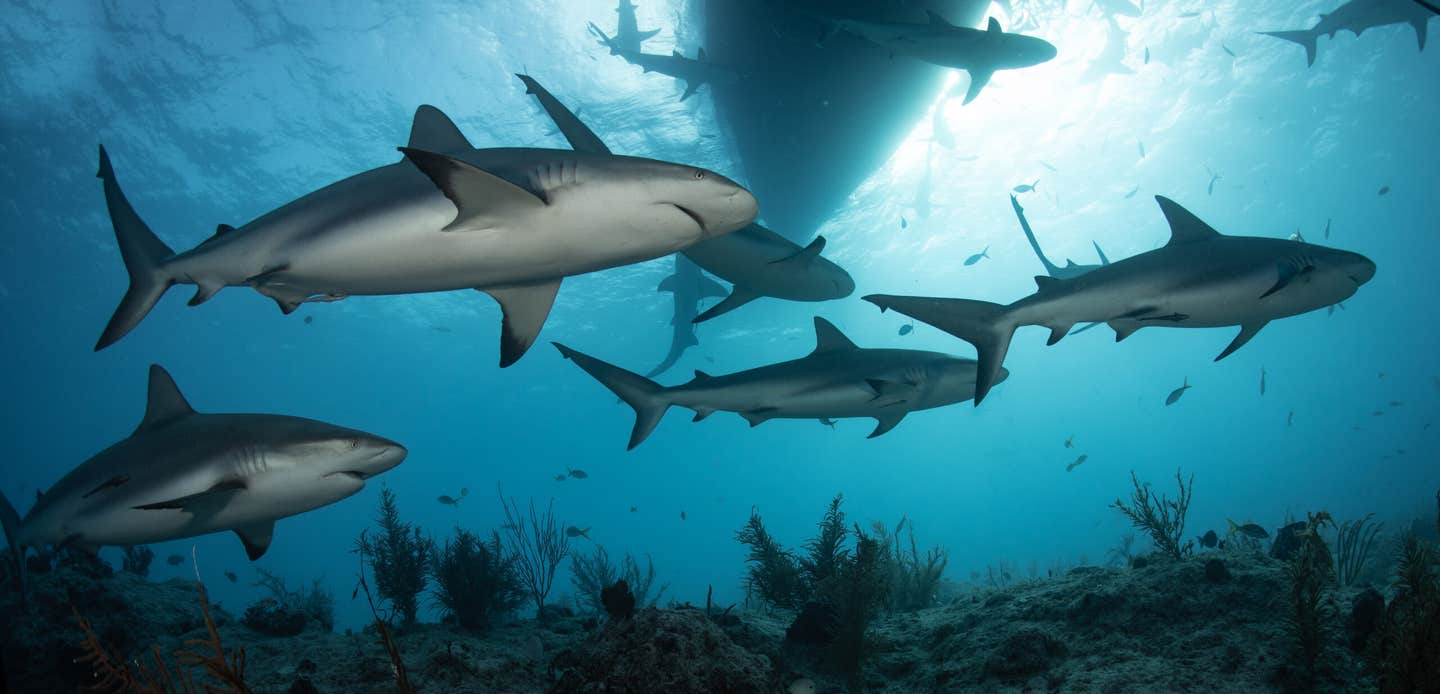
Micro-CT scans uncovered how six shark species carry spinal designs tuned to their distinct swimming styles. (CREDIT: Shutterstock)
A shark’s spine looks simple from the outside. Inside, however, it carries a carefully arranged network of mineral plates and branches.
That hidden structure helps determine whether a shark races through open water, turns sharply, or swings an oversized tail at prey.
Florida Atlantic University researchers and NOAA Fisheries collaborators examined the vertebrae of six shark species.
The team studied great white, shortfin mako, porbeagle, common thresher, sand tiger and basking sharks. Each species uses its body differently while swimming. The analysis found no single vertebral design shared across every shark. Instead, each spine matched the forces created by its swimming style.
“By examining species with varied swimming strategies, we found that the shark spine is not a one-size-fits-all design,” said Jamie Knaub.
“Instead, its internal architecture reflects the unique demands of each species’ movement.”
Knaub is the study’s first author and a research specialist at FAU Laboratory Schools. She is also a doctoral candidate in FAU’s Department of Biology.
Looking Beneath a Cartilage Skeleton
Sharks have relied on cartilage-based skeletons for more than 400 million years. Their spines must withstand millions of repeated swimming motions.
Cartilage gives sharks flexibility without the weight of a fully bony skeleton. Yet some parts of each vertebra contain mineralized tissue. Those reinforced areas can stiffen the spine and help it resist heavy loads. Their placement matters as much as their quantity.
Scientists already understood much about shark muscles and swimming movements. The inner design of the spine remained far less clear.
The research team collected vertebrae from several points along each shark’s body. These included front, middle and rear sections. High-resolution micro-computed tomography created three-dimensional images without cutting or damaging the samples.
“High-resolution micro-CT imaging gave us a window into the hidden world inside shark vertebrae,” Knaub said.
The scans exposed structures shaped across hundreds of millions of years of evolution. They also allowed detailed comparisons among species.
Knaub scanned the vertebrae at the Berlin Family Bioimaging Lab. The laboratory sits within FAU Lab Schools’ Marcus Research and Innovation Center.
The team measured each vertebra’s size and shape. Researchers also mapped mineralized plates and branching internal structures. They then compared those features along the spine and among sharks with very different swimming habits.
The Spine Changes from Front to Tail
The middle section consistently contained the largest vertebrae. That area may serve as a hinge within the body wave created during swimming.
Vertebrae near the tail faced a different mechanical challenge. They had to manage strong forces during repeated tail beats. In fast-swimming sharks, rear vertebrae contained more mineralized plates. That extra reinforcement likely increased stiffness near the tail.
A stiffer rear spine can transfer muscular energy more efficiently. More of that energy then reaches the tail and drives propulsion.
The pattern appeared in the great white, shortfin mako and porbeagle. All three move quickly through open water.
Their vertebral columns combined large middle sections with reinforced rear sections. This arrangement supports stability and efficient energy transfer. Closely related species also shared similar designs. Great white and shortfin mako vertebrae appeared especially alike.
That similarity may reflect both family relationships and comparable swimming mechanics.
The sand tiger shark followed a different plan. Its vertebrae likely allow greater flexibility through more of the body. That design suits a shark that swims more slowly and maneuvers through complex underwater spaces.
“The shark spine isn’t simply flexible,” said senior author Marianne E. Porter from FAU’s Department of Biological Sciences. “It’s optimized to balance strength, stiffness and motion in ways that maximize performance.”
A Tail Built for Striking
The common thresher shark produced one of the study’s most striking patterns. This species carries an exceptionally long tail. It uses that tail in powerful movements that can stun prey.
Its vertebrae contained the greatest quantity of mineralized plates and branching structures among the sharks examined. Those reinforcements appear suited for repeated sideways motion and overhead tail strikes.
The thresher’s spine must support ordinary swimming while surviving forces created during hunting. That requires both strength and controlled motion. Its internal architecture differs sharply from species that simply push water backward with more conventional tail beats.
The basking shark stood at the opposite end of the range. It is the world’s second-largest fish, yet it moves slowly while filtering food from seawater.
Its vertebrae had far less mineralization than those of the faster predators. That reduced reinforcement reflects a different set of forces. A slow filter feeder does not need the same stiff energy-transfer system.
The contrast between thresher and basking sharks was especially clear. Both are large animals, but size alone did not dictate spinal design. Movement mattered.
Images Turn Anatomy into Mechanics
The micro-CT scans did more than display unusual shapes. They helped connect internal anatomy with likely physical function.
“This work would not have been possible without advanced micro-CT technology,” said co-author Tricia L. Meredith.
The method exposed three-dimensional structures that researchers could not easily access before. “These imaging capabilities allow us to move beyond simply describing anatomy to understanding how internal structures function mechanically,” Meredith said.
Meredith directs research for A.D. Henderson University School and FAU High School. She is also an assistant research professor. The research connects the position of mineral tissue with swimming needs. However, it does not turn each feature into a final mechanical answer.
The study linked patterns with known movement styles across the six species. Those links provide a base for deeper mechanical testing.
The findings also underscore the value of studying multiple body regions. A single vertebra cannot describe the entire spine. The front, middle and tail regions experience different loads during swimming.
Those local demands shaped the size, form and reinforcement of the vertebrae.
Nature did not build one shark spine and reuse it unchanged.
From Ancient Predators to New Materials
The research may interest engineers as well as biologists.
Shark vertebrae combine lightweight cartilage with carefully placed mineral reinforcement. That balance creates strength without making the whole spine rigid. Human-made systems often face the same tradeoff.
Robotic parts may need to bend while resisting repeated stress. Biomedical devices may require softness in one area and stiffness elsewhere. The shark spine offers several natural examples of that balance.
“Nature has spent hundreds of millions of years refining these designs,” Porter said.
Engineers could study how mineral plates change across a flexible framework. Similar strategies might guide materials that adjust strength by location.
The researchers pointed to possible uses in robotics and biomedical technology.
Such applications remain future possibilities. The current work focused on comparative anatomy and shark movement.
Study co-authors included FAU biological sciences undergraduates Madisan Biordi and Emma Pawlik. Michelle Passerotti, leader of the NOAA Fisheries Apex Predators Program, also contributed. Lisa J. Natanson, is a researcher at NOAA Fisheries, and served as another co-author.
Practical Implications of the Research
The findings give scientists a clearer way to connect shark anatomy with swimming behavior. Researchers can now compare where vertebrae become larger, shorter, more flexible or more reinforced along the body.
That knowledge may improve future studies of shark movement, feeding and ecological adaptation. It could also help scientists interpret why related species share certain spinal traits while distant species differ.
For engineers, the work offers a model for building light structures with targeted reinforcement. Instead of making an entire material stiff, designers could strengthen only areas that face the greatest forces.
That approach may support flexible robots, durable implants or other devices that must bend without failing.
The study also highlights a wider lesson from animal anatomy. A skeleton does not simply hold the body together. Its internal design can preserve energy, direct motion and support specialized behavior.
In sharks, those adaptations sit beneath the surface, hidden inside a spine built for a particular way of moving.
Research findings are available online in the journal Journal of Anatomy.
The original story "FAU scientists discover that shark spines are not one-size-fits-all" is published in The Brighter Side of News.
Related Stories
- Deep-ocean cameras capture first-ever images of live Goblin shark
- Whale fossils reveal evidence of ancient shark attacks
- Bull sharks have friends, and they choose them carefully
Like these kind of feel good stories? Get The Brighter Side of News' newsletter.
Hannah Shavit-Weiner
Medical & Health Writer
Hannah Shavit-Weiner is a Los Angeles–based medical and health journalist for The Brighter Side of News, an online publication focused on uplifting, transformative stories from around the globe. Having published articles on AOL.com, MSN and Yahoo News, Hannah covers a broad spectrum of topics—from medical breakthroughs and health information to animal science. With a talent for making complex science clear and compelling, she connects readers to the advancements shaping a brighter, more hopeful future.