Lab-grown mini lenses offer a closer look at how cataracts develop
Scientists grew human lens organoids with specialized proteins and organized fibers, creating a model to study cataracts and explore future regenerative care.
 Anglia Ruskin University Writer: Jamie Forsyth

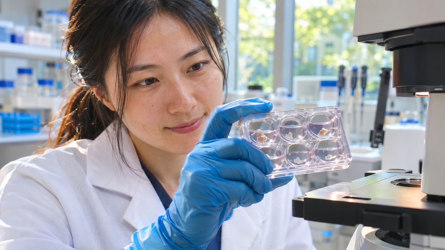
A researcher examines cultured tissue in this illustrative laboratory scene. Scientists grew human lens organoids that reproduce key features of the eye’s lens, offering a new model for cataract research. (CREDIT: The Brighter Side of News)
- Human stem cells formed miniature lens-like tissues with organized fibers, lens proteins and a capsule-like covering.
- A specialized growth medium improved tissue development, while cells taken from the structures produced secondary lens-like clusters.
- The findings offer a model for cataract research, but do not establish a replacement lens or a treatment for patients.
Tiny clusters of human cells have developed several features that help an eye lens stay clear. Grown in laboratory dishes, these miniature structures contain specialized proteins, elongated fibers and a protective capsule-like boundary. They give scientists a closer view of how human eye tissue develops, and how that process might go wrong.
The work brings together researchers from Anglia Ruskin University, The University of Osaka and Cardiff University. Their findings appear in npj Regenerative Medicine. The team developed an improved way to grow lens organoids, small tissue models that reproduce selected features of a natural lens.
Those models could help researchers investigate cataracts, the clouding that prevents a lens from focusing light properly. The World Health Organization estimates that cataracts account for 94 million cases of distance vision impairment or blindness within its estimate of preventable or unaddressed vision loss. The experiment did not test a cataract treatment in patients or demonstrate a transplant-ready lens.
Barbara Pierscionek, a professor at ARU's Centre for Biomedicine and Health Innovation, said: “This is a major step forward as it is very difficult to obtain and study human eye lens tissue while it is developing. We've now successfully grown miniature human lens tissue from stem cells in the laboratory and identified the conditions needed for it to closely resemble a natural human lens.”
Why a clear lens is difficult to build
A natural lens must do more than let light pass through it. Its carefully arranged cells and proteins help with focusing light onto the retina, the light-sensitive tissue at the back of the eye. Reproducing that architecture requires several kinds of cells to develop and organize together, rather than simply making a transparent-looking cluster.
The lens has three major components: an outer capsule, a thin layer of epithelial cells and elongated fiber cells. Epithelial cells support growth, while mature fibers form much of the lens interior. As fibers mature, they remove their nuclei and other internal structures, reducing obstacles that could scatter incoming light.
Proteins called crystallins are central to this arrangement. They remain densely packed and soluble, helping maintain transparency and the lens's ability to bend light. Some crystallins also act as molecular chaperones, helping protect other proteins from damage. Understanding that protein quality control is important because mature lens fibers cannot continually replace their proteins.
When proteins become damaged or aggregate, the lens can lose its clarity. Scientists therefore need models that capture both tissue organization and the molecules responsible for keeping it transparent. Developing human lens tissue is difficult to obtain, making it hard to study these processes directly or follow them over time.
Growing human lens organoids from stem cells
The researchers began with human induced pluripotent stem cells, or iPSCs. These are adult cells reprogrammed into a state capable of producing many cell types. Such cellular reprogramming creates a starting material for studying development without obtaining tissue from a developing human eye. This study used an established human iPSC line called 201B7.
The team used an approach that lets cells organize into concentric regions associated with different eye tissues. It then compared a standard eye-development medium with a lens-optimized medium, a nutrient and signaling mixture designed to encourage lens formation. This builds on the broader use of organoids to examine how cells organize into tissue.
Earlier research in npj Regenerative Medicine also produced patient-specific lens-like structures that modeled inherited cataracts, showing that the present advance builds on an existing field.
Across eight weeks, the two growth conditions produced different patterns. The optimized medium encouraged an expanded lens-associated region and larger, more frequent lens-like bodies, known as lentoids. Under standard conditions, the tissue retained more features associated with epithelial cells and showed less advanced fiber organization. The comparison connected changes in the culture environment with changes in tissue development.
Gene measurements supplied another line of evidence. The optimized cultures showed stronger activity in genes encoding several crystallins, especially gamma-crystallins associated with later fiber development. Certain gamma-crystallin genes showed increases of roughly 21-fold to 63-fold compared with the standard medium. These were changes in gene expression, not measurements showing an equivalent improvement in vision or transparency.
Looking inside the miniature structures
The researchers combined gene analysis with protein staining and microscopy. Staining helped establish where different proteins appeared, while electron microscopes revealed much finer structural details. Together, these methods tested whether the cultures contained organized lens-like tissue rather than merely cells producing a few characteristic proteins.
The optimized cultures contained elongated fiber-like cells arranged in layers. Researchers also found membrane junctions and interlocking features resembling the ball-and-socket connections seen between natural lens fibers. Proteins involved in cell adhesion and communication appeared together within the lentoids. Their locations supported the interpretation that the cells were developing coordinated lens-like architecture.
At the outside, the team detected collagen IV and perlecan, components of the lens capsule's supporting matrix. These proteins surrounded the optimized lentoids, complementing the evidence for fiber-like and epithelial-like cells. Reproducing all three major structural components strengthens the model's resemblance to human lens tissue. It does not establish that the structures perform every task of a natural lens.
Some questions about maturation remain. Electron microscopy showed fibers without obvious nuclei or organelles, but nuclear staining persisted in parts of the cultures. The researchers noted that nearby or surrounding cells complicate interpretation, and complete nuclear clearance may require different growth conditions. Measurements of refractive index, which describes how strongly material bends light, also remain a future step.
A research model with regenerative potential
Cells taken from the lentoids offered an additional finding. When researchers placed the structures onto a supportive surface, epithelial-like cells spread outward and could subsequently form secondary lens-like structures. Their capacity to expand was limited, however. This suggests regenerative potential within the culture system, rather than proof that an adult eye could regrow its lens.
The result could help scientists investigate how lens cells retain developmental flexibility. It also adds a laboratory tool to the wider search for regenerative eye treatments. Researchers still need to trace cell origins more precisely and establish how reliably these populations can be maintained. Future work could explore patient-specific cells or gene editing to model inherited cataracts.
The platform could also support drug testing, allowing scientists to examine how candidate compounds affect developing lens tissue. Pierscionek described that possibility cautiously. “As the technology develops, these lab-grown tissues could help us test new medicines, better understand diseases that affect vision, and potentially contribute to the development of future regenerative treatments.”
For now, surgery remains the established treatment for removing cataracts; these cultures offer a research system, not an alternative available to patients. Future experiments must connect their promising cellular organization with measured optical performance. A central question is whether more mature lab-grown tissue can reproduce the transparency and light-bending properties that make a natural lens work.
Dig deeper into lens development and cataracts
These resources explain cataract care and earlier approaches to studying or regenerating lens tissue.
Cataracts
Describes cataract symptoms, diagnosis and established treatment options for patients. (National Eye Institute, updated August 2026)
Blindness and vision impairment
Explains the global burden of cataracts and other causes of vision impairment, including gaps in access to care. (World Health Organization, February 2026)
Lens regeneration in situ using hESCs-derived cells -similar to natural lens
Examines lens regeneration using human embryonic stem-cell-derived cells in an experimental animal setting. (iScience, May 2023)
Modeling congenital cataract in vitro using patient-specific induced pluripotent stem cells
Shows how patient-derived lens-like tissues can reproduce features of inherited cataracts and support laboratory drug testing. (npj Regenerative Medicine, October 2021)
Lens regeneration using endogenous stem cells with gain of visual function
Investigates a different regenerative strategy that preserves a patient's own lens epithelial cells during cataract surgery. (Nature, March 2016)
Research findings are available online in the journal npj Regenerative Medicine.
The original story "Lab-grown mini lenses offer a closer look at how cataracts develop" is published in The Brighter Side of News.
Related Stories
- Lifechanging cataracts treatment eliminates the need for surgery
- Spiral-shaped lenses revolutionize cataracts and vision treatment
- Smart glasses turn blinks and jaw movements into precise health monitoring
Like these kind of feel good stories? Get The Brighter Side of News' newsletter.
Mac Oliveau
Writer
Mac Oliveau is a Los Angeles–based science and technology journalist for The Brighter Side of News, an online publication focused on uplifting, transformative stories from around the globe. Having published articles on MSN, and Yahoo News, Mac covers a broad spectrum of topics including medical breakthroughs, health and green tech. With a talent for making complex science clear and compelling, they connect readers to the advancements shaping a brighter, more hopeful future.