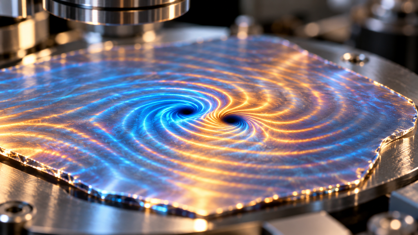
Miniature brain and cardiovascular models head to orbit on SpaceX CRS-35 to study aging
Brain and cardiovascular models aboard CRS-35 will probe how spaceflight affects living human tissue and aging-related biology.
 Mount Sinai Writer: Stacy Anderson

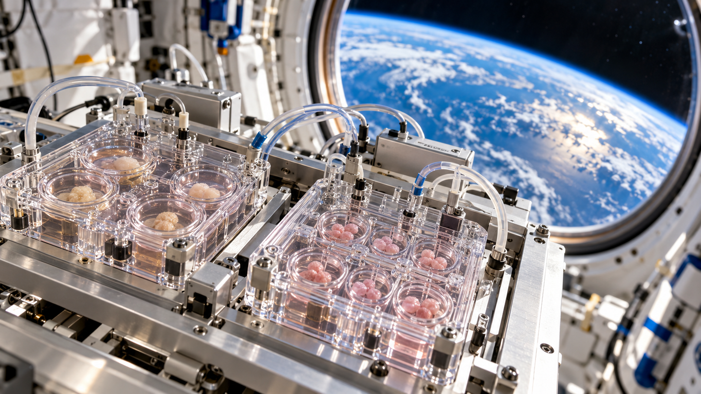
Mount Sinai sends brain and heart tissue models to the ISS to study spaceflight, genetic risk and changes linked to aging. (CREDIT: The Brighter Side of News)
- Mount Sinai brain and cardiovascular tissue models launched toward the International Space Station aboard SpaceX CRS-35.
- The experiments will examine tissue function, genetic susceptibility and biological changes associated with spaceflight.
- Published miBrain research and a separate circulation preprint provide the groundwork; results from this mission remain ahead.
Human brain tissue and beating cardiovascular organoids are headed into orbit to face an unusual laboratory test. Their destination is the International Space Station, where weightlessness and radiation exposure could reveal vulnerabilities difficult to study on Earth.
The Mount Sinai investigations are traveling aboard SpaceX CRS-35, which launched October 13. They bring together researchers in stem cell biology, neuroscience, surgery and cardiovascular science at the Icahn School of Medicine.
One project uses a personalized brain tissue model called miBrain. The other places cardiovascular organoids inside microfluidic chips, miniature devices that manage fluid around living tissue.
The mission builds on established laboratory work, including a 2025 paper in PNAS introducing miBrain. However, the new flight is an experiment in progress. Its findings have yet to establish how these particular models respond to space.
A brain model built from six cell types
Neurons rarely work alone. They communicate within a network of supporting cells, immune cells and blood vessels, whose interactions help maintain brain health.
The miBrain platform brings six major cell types into a three-dimensional tissue culture. These include neurons, astrocytes, microglia, oligodendrocytes, pericytes and endothelial cells.
Each plays a different role. Neurons transmit signals, while oligodendrocytes produce myelin, the insulating material around nerve fibers. Microglia provide immune functions, and endothelial cells help form vessels.
Developed through work involving MIT professors Li-Huei Tsai and Robert Langer, alongside Joel Blanchard, miBrain uses an engineered hydrogel. That material supports the cells as they assemble into interacting networks.
The published research documented neuronal activity, vascular barrier features and engagement between myelin-producing cells and neurons. These capabilities make the culture more representative of brain tissue than isolated cells growing alone.
Still, reproducing selected functions does not recreate an entire brain. The model offers access to specific cellular relationships that scientists can measure and manipulate.
Patient-derived stem cells add another advantage. A culture can carry a donor’s genome, allowing researchers to examine how inherited differences affect tissue behavior. Gene editing can also produce closely matched cultures with different versions of a selected gene.
Testing neurological risk in orbit
Blanchard, an associate professor at Mount Sinai, leads the brain investigation. Working with NASA and Space Tango, his team plans a series of flights to assess brain cell signaling and function.
The research will examine why responses to spaceflight may differ among people. It will also explore how miBrain changes with age and how genetic risks influence that process.
One target is APOE4, a gene variant associated with increased risk of Alzheimer’s disease. Comparing tissue with different APOE variants could help separate inherited susceptibility from effects shared across the models.
“Our research could establish miBrain as a practical tool for assessing neurological risk before and during spaceflight,” Blanchard said.
His team aims to identify biomarkers of injury, assess genetic susceptibility and evaluate protective countermeasures. Those are research goals, rather than capabilities already demonstrated by the current mission.
The distinction matters because spaceflight changes several conditions together. Microgravity, radiation exposure and the demands of keeping tissue alive in flight can complicate interpretation.
A tissue model provides a controlled starting point for investigating mechanisms. Connecting those mechanisms to an astronaut’s health, or to aging on Earth, will require further evidence.
Beating cardiovascular tissue travels alongside it
The cardiovascular investigation uses healthy human stem cells to grow beating organoids. They sit inside a microfluidic device called the µCVchip.
Two chips containing up to 16 cardiovascular organoids are traveling in Space Tango’s automated CubeLab system. The equipment will perform daily electrical pacing, pressure testing, microscopic imaging and fluid sample collection.
Together, these measurements should help researchers track cardiac and vascular function during extended microgravity exposure. Automation allows repeated observations without relying on constant hands-on work by astronauts.
Kevin Costa and David Sachs, both faculty members in Mount Sinai’s Department of Surgery, collaborate on the project. They hope the findings will guide ways to protect astronauts’ hearts and inform research into cardiovascular disease.
The team previously sent stem cell-derived cardiac muscle cells into space on CRS-29 in 2023. That earlier work assessed cell viability. The current flight advances to more complex cardiovascular models and physiological measurements.
Spaceflight can produce cardiovascular changes resembling some effects of aging. Studying those changes in engineered tissue may expose useful biological pathways, although similarities do not make spaceflight identical to ordinary aging.
Circulation adds another layer of complexity
A separate 2025 preprint describes a vascular-perfusable brain-on-chip related to miBrain. It reports laboratory work on Earth, not findings from the new space mission.
The researchers developed a microfluidic platform called GelChip to support vessel formation within an engineered hydrogel. Alongside six brain cell types, the cultures included human fibroblasts.
Device dimensions proved important. Larger chamber heights supported more connected vascular networks, helping overcome problems created by hydrogel swelling in confined spaces.
After connecting the networks to inlet and outlet ports, the team demonstrated tracer movement through small vessels. The approach provides a way to investigate transport across a modeled blood-brain barrier.
That barrier controls what passes from circulation into brain tissue, protecting the brain while complicating drug delivery. A model with accessible vessels could help investigators examine both sides of that problem. They could study changes in barrier properties and test how candidate compounds move through the engineered system.
APOE4 cultures showed vascular differences compared with closely matched APOE3 cultures. Replacing individual cell types also implicated APOE4 astrocytes in reduced tight-junction marker signals.
These observations remain preliminary. The study used one matched stem-cell pair for its genetic comparisons, and the authors called for testing additional lines. Perfusion also became less reliable as cultures included more cell types.
What the flight can establish
The immediate challenge is to learn whether these complex human tissue systems remain useful and measurable in orbit. Reliable measurements would support later investigations into injury, inherited risk and possible protections.
Their value comes from examining human cells within interacting tissue environments. That offers a different perspective from studying individual cells, animals or astronauts alone.
Future medical applications will depend on whether findings hold across donors, experiments and relevant clinical settings. Neither project has yet demonstrated an antiaging treatment or a way to predict an individual astronaut’s outcome.
For now, the flight extends two laboratory platforms into an environment that can stress biological systems differently. The next step is to see what those living models record.
Dig deeper into human tissue research in space
These resources explain earlier experiments and the broader rationale for studying engineered tissues in orbit.
- Spaceflight-induced contractile and mitochondrial dysfunction in an automated heart-on-a-chip platform reports functional changes in engineered heart tissue after spaceflight. (PNAS, 2024)
- Effects of microgravity on human iPSC-derived neural organoids on the International Space Station examines earlier neural organoid experiments in orbit. (Stem Cells Translational Medicine, 2024)
- Mount Sinai Bioengineers Send Cardiac Muscle Samples Into Space to Study Heart Cell Biology in Microgravity describes the team’s earlier cardiac cell mission. (Mount Sinai, 2023)
- Tissue Chips Accurately Model Organs in Space introduces tissue-chip research aboard the station. (NASA, 2024)
- NASA’s Twins Study Results Published explains findings from comparing an astronaut with his Earth-based identical twin. (NASA, 2019)
Research findings are available online in the journal PNAS.
The original story "Miniature brain and cardiovascular models head to orbit on SpaceX CRS-35 to study aging" is published in The Brighter Side of News.
Related Stories
- Space travel accelerates stem cell aging - threatening astronaut health
- MIT researchers use light to identify aging cells without destroying them
- New CPR simulator could help save astronauts in space
Like these kind of feel good stories? Get The Brighter Side of News' newsletter.
Joseph Shavit
Writer, Editor-At-Large and Publisher
Joseph Shavit, based in Los Angeles, is a seasoned science journalist, editor and co-founder of The Brighter Side of News, where he transforms complex discoveries into clear, engaging stories for general readers. With vast experience at major media companies like The Los Angeles Times, Times Mirror and Tribune Publishing, he writes with both authority and curiosity. His writing focuses on space science, planetary science, quantum mechanics, geology. Known for linking breakthroughs to real-world markets, he highlights how research transitions into products and industries that shape daily life.