
MIT researchers use light to identify aging cells without destroying them
Light-based chemical fingerprints could eventually help scientists find aging cells inside tissue without conventional destructive tests.
 MIT Writer: Anne Trafton

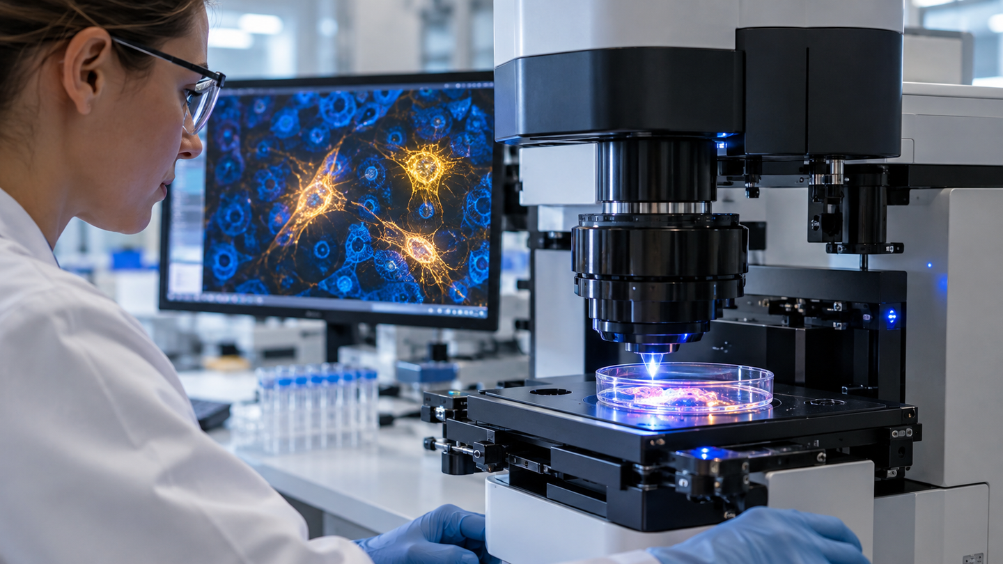
MIT researchers use Raman microscopy and gene data to identify biochemical barcodes of senescent cells without destroying tissue. (CREDIT: Shutterstock)
- MIT researchers combined Raman microscopy with single-cell and spatial gene-expression measurements to identify biochemical patterns associated with cellular senescence without destroying the tissue being examined.
- Senescent cells showed distinctive molecular changes, including a recurring lipid-associated Raman signature, while lung and skin displayed different age-related gene programs.
- The team used these combined signals to build a machine-learning “barcode” for senescence, raising the possibility of future imaging tools that could locate senescent cells inside living tissue.
Cells that stop dividing but refuse to disappear are notoriously difficult to identify without destroying them. MIT researchers have now developed an optical approach that could eventually allow scientists to find these senescent cells while leaving surrounding tissue intact.
The method combines Raman microscopy, which reads the chemical fingerprints of molecules using light, with detailed measurements of gene activity. Researchers used those complementary views to identify combinations of biochemical and genetic features associated with senescence.
The resulting platform, called RamanOmics, was tested in lung and skin tissue from young and old mice. The study, published in Nature Aging, found that senescent cells carry recognizable biochemical signatures that can be translated into a machine-learning “barcode.”
The work remains preclinical, but researchers envision a future in which Raman-based instruments could locate senescent cells without first removing or destroying them.
“You can imagine that one day we may develop an endoscope that can look inside your body and identify cellular senescence,” senior author Jeon Woong Kang said.
Senescent cells are difficult to define with one marker
Cellular senescence often begins after DNA damage or other stresses cause a cell to permanently stop dividing. The cell remains metabolically active, however, and can undergo major changes in shape, gene activity and the molecules it releases.
Senescence is not inherently harmful. These cells have important roles during development, wound repair and tissue regeneration. Problems can emerge when they persist and accumulate, particularly with age.
The immune system normally helps remove unwanted senescent cells. That clearance can become less effective over time, allowing populations of senescent cells to remain in tissues and contribute to chronic inflammation, fibrosis and age-associated diseases.
Scientists often identify them using markers such as p16 or p21, proteins associated with cell-cycle arrest. No single biomarker captures every form of senescence, however, and many conventional techniques require cells to be fixed, stained or destroyed.
The NIH Cellular Senescence Network, or SenNet, is addressing that problem by developing maps, definitions and technologies for identifying different senescent cell states throughout the body.
Raman microscopy reads chemistry without destroying cells
Raman microscopy works by shining light onto a sample and measuring tiny shifts in the light scattered back.
Those shifts reflect molecular vibrations associated with chemical bonds. A Raman spectrum can therefore contain information about lipids, proteins, nucleic acids, sugars and other cellular components without requiring fluorescent labels or destructive chemical processing.
The technique provides a very different view from RNA sequencing. Gene-expression measurements reveal which genes cells are using, while Raman measurements reveal aspects of the biochemical state those genetic programs help create.
Researchers had previously demonstrated that Raman images could be used to predict RNA-expression profiles in live cells. The earlier Raman2RNA technique showed that optical biochemical information and gene-expression states are closely related.
The new study extended that idea into intact tissue. Researchers collected lung and skin samples from 2-month-old and 26-month-old mice and combined hyperspectral Raman imaging with single-nucleus RNA sequencing and STARmap spatial transcriptomics.
This allowed them to examine the chemistry and gene activity of individual cells while preserving information about where those cells were located within tissue.
Aging left different signatures in lung and skin
The experiments showed that aging does not produce one universal molecular pattern across tissues.
In older lung tissue, researchers found increased activity in pathways associated with immune activation and inflammation. Endothelial cells and other populations also showed changes connected with extracellular matrix remodeling.
Old skin followed a different trajectory. Researchers found altered metabolic and structural programs, including changes involving keratinocytes, fibroblasts, collagen and other components of the tissue environment.
Senescent cells themselves also differed according to tissue and age. Older lung cells showed signatures consistent with fibrosis and inflammation, while younger senescent cells retained stronger patterns associated with repair and DNA-damage responses.
That distinction supports a growing view of senescence as a collection of cellular states rather than a single biological condition. NIH researchers have recently proposed the term “senotypes” to describe distinct forms shaped by tissue, age and physiological context.
Lipids emerged as an important biochemical clue
Raman imaging added information that gene expression alone could not reveal.
Researchers identified dozens to hundreds of spectral differences between senescent and nonsenescent cells, depending on tissue and cell type. Some signals corresponded to proteins, nucleic acids, sugars and collagen.
One of the clearest recurring changes involved lipids.
Raman peaks around 1,131 to 1,135 inverse centimeters were elevated in p21-positive senescent cells across tissues. The researchers linked those spectral changes to lipid-associated molecular structures. The published study identifies this lipid signature as one of the biochemical features conserved across mouse lung and skin.
The reason senescent cells accumulate or reorganize particular lipids remains uncertain. Lipid metabolism affects membranes, signaling and inflammatory responses, giving researchers several possible mechanisms to investigate.
Other changes remained tissue specific. Senescent skin cells showed alterations related to collagen and epidermal differentiation, while lung cells displayed different nucleic-acid and protein-associated signals.
Machine learning combines the clues into a barcode
The researchers next asked whether those biochemical signals could help identify senescent cells automatically.
They combined selected Raman peaks with gene-expression features and trained a random-forest machine-learning classifier. Using both forms of information improved classification compared with gene-expression information alone.
The team then ranked the most informative features and organized them into multimodal barcodes representing senescent and nonsenescent states.
The goal is not necessarily to perform RNA sequencing every time a tissue is examined. Instead, gene-expression measurements can help researchers establish which Raman signals correspond to senescence, after which a smaller set of optical signatures might eventually be used for faster detection.
“Using this barcode, we can focus on a few Raman bands that emerged as the most informative in this work,” study author Salvatore Sorrentino said.
The current imaging system is not yet fast enough for routine medical use. Analyzing a tissue region approximately one square millimeter in area currently takes around 30 hours, so the team is developing faster instruments focused on the most informative wavelengths.
Human testing is the next major hurdle
The study was performed in mouse tissues, and researchers cannot yet assume the same Raman barcodes will accurately identify senescence in people.
Human tissues are more heterogeneous, and senescent cells can differ depending on disease, organ, age and the stress that caused them to stop dividing. Any future diagnostic technology will require extensive validation across human samples.
Still, a nondestructive technique could address a fundamental weakness in current senescence research. Scientists would potentially be able to locate and monitor cells while preserving the tissue for further study.
That ability could become particularly useful as researchers test senolytics and other therapies intended to eliminate or modify harmful senescent cells. Rather than relying only on destructive biopsies and molecular assays, future Raman instruments might provide a direct optical readout of whether the cellular landscape has changed.
The immediate accomplishment is more modest but important. Senescent cells do not differ only in the genes they express. Their underlying chemistry also leaves measurable fingerprints, and light may provide a way to read them.
Dig deeper into cellular senescence and Raman imaging
These resources provide additional context on senescent-cell mapping, nondestructive molecular imaging and efforts to distinguish different senescent states.
Prediction of single-cell RNA expression profiles in live cells by Raman microscopy with Raman2RNA: This earlier work demonstrated that Raman images can predict single-cell gene-expression profiles without destroying living cells. (Nature Biotechnology, 2024)
Cellular Senescence Network (SenNet): The NIH program is building comprehensive maps and technologies for identifying senescent cells across tissues and stages of life. (National Institutes of Health, ongoing)
Senescent cells mapped in human body over the lifespan: This NIH overview describes recent efforts to classify and map senescent cells in human tissues. (National Institutes of Health, 2026)
Vibrational spectroscopic imaging of living systems: An emerging platform for biology and medicine: This review explains how Raman and related vibrational methods reveal biochemical information from living cells and tissues. (Science, 2015)
NIH SenNet Consortium to map senescent cells throughout the human lifespan to understand physiological health: This consortium paper lays out the strategy for defining senescence across tissues rather than relying on a single universal biomarker. (Nature Aging, 2022)
Research findings are available online in the journal Nature Aging.
The original story "MIT researchers use light to identify aging cells without destroying them" is published in The Brighter Side of News.
Related Stories
- New optical shutter uses light and AI to see through dense fog and tissue
- New light-activated heart tissue revolutionizes heart repair
- Global first: AI tool predicts how proteins work in cells and tissues
Like these kind of feel good stories? Get The Brighter Side of News' newsletter.
Rebecca Shavit
Writer
Based in Los Angeles, Rebecca Shavit is a dedicated science and technology journalist who writes for The Brighter Side of News, an online publication committed to highlighting positive and transformative stories from around the world. Having published articles on MSN, AOL News, and Yahoo News, Rebecca's reporting spans a wide range of topics, from cutting-edge medical breakthroughs to historical discoveries and innovations. With a keen ability to translate complex concepts into engaging and accessible stories, she makes science and innovation relatable to a broad audience.