Texas A&M study finds humans may have hidden regenerative powers
A two-step treatment in mice redirected wound healing from scarring toward regrowth of bone, joints, and ligaments.

 Edited By: Joseph Shavit
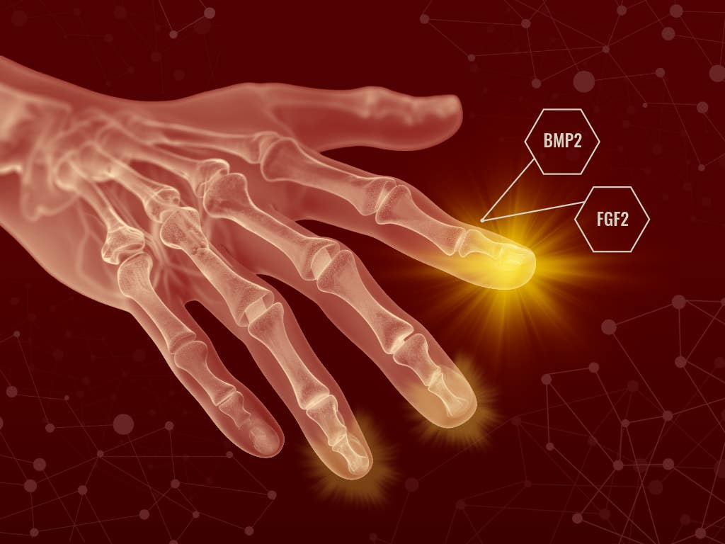
A conceptual graphic shows how growth factors BMP2 and FGF2 are applied to the injury site to stimulate tissue regeneration, highlighting new research into restoring damaged digits. (CREDIT: Melissa Bristow/Texas A&M University College of Veterinary Medicine and Biomedical Sciences)
A mouse digit does not usually grow back once it is amputated above the tip. It seals the wound, lays down scar tissue, and stops there. That familiar limit has shaped how scientists think about healing in mammals, and about why humans do so poorly at replacing what injury takes away.
A new study from Texas A&M suggests that limit may be less absolute than it looks.
By treating injured mouse digits with two growth factors in sequence, researchers were able to trigger the regrowth of an anatomically complete, though imperfect, set of structures at the amputation site. The response included bone, joint tissue, ligaments, tendons, and a blastema-like mass of cells, the kind of regeneration-linked structure better known from salamanders than mammals.
“Why some animals can regenerate and others, particularly humans, can't is a big question that has been asked since Aristotle,” said Dr. Ken Muneoka, a professor in the Department of Veterinary Physiology & Pharmacology at the Texas A&M College of Veterinary Medicine and Biomedical Sciences. “I've spent my career trying to understand that.”
A fork in the healing process
The work, published in Nature Communications, focuses on what happens after amputation in neonatal mice at a level known to be non-regenerative. Instead of rebuilding the missing part, the wound normally heals with fibrosis. Fibroblasts close the injury quickly, but the repair produces scar tissue rather than new anatomy.
Animals that regenerate take a different route. In salamanders, cells gather into a blastema, a temporary cluster that can drive the regrowth of amputated structures. Muneoka and his colleagues wanted to know whether mammalian wound cells might still be capable of entering a similar state.
“It’s as if these cells can move in two different directions,” Muneoka said. “They could either make a scar or make a blastema. Our research focused on redirecting the behavior of fibroblasts already present at the injury site.”
The team used a two-step treatment. After the wound had already closed, they first applied fibroblast growth factor 2, or FGF2. Several days later, they applied bone morphogenetic protein 2, known as BMP2.
The sequence mattered. FGF2 alone caused wound cells to accumulate into a proliferating, blastema-like outgrowth in the dorsal part of the amputation site. It also triggered a separate ventral response tied to a forming joint cavity, but by itself it rarely produced substantial regeneration. When BMP2 was added after that first step, the result was much stronger.
“This is really a two-step process,” Muneoka said. “You first shift the cells away from scarring, and then you provide the signals that tell them what to build.”
An incomplete digit, but a striking result
The researchers found that every digit treated with the FGF2-to-BMP2 sequence formed ectopic bone by the end of the experiment. Most regenerated one or two new skeletal elements. Their analysis showed that one commonly formed in a dorsal position and resembled the missing distal phalanx, while another often formed in a distal-ventral position and resembled a sesamoid bone.
The structures were not exact copies of the originals. Even so, they were not random bits of tissue either.
Histology showed a vascularized marrow cavity in the phalanx-like element, tendon and ligament attachments, articular cartilage in many samples, and in some cases a newly formed ligament linking the regenerated elements. The team concluded that the treatment had produced a complete but imperfect digit regenerate.
“We regenerated what you would expect to see at that level of injury,” Muneoka said. “The structures are there, just not in a perfect form.”
The study also draws a distinction between two repair modes happening in the same wound. One path depended on the blastema-like structure and led to regeneration of the distal skeletal element. The other did not appear to require a blastema and instead rebuilt a synovial joint complex, including connective tissues.
That split, the authors argue, points to a wound environment that can steer cells toward very different outcomes, from fibrosis to partial regeneration to something closer to epimorphic regeneration, the process seen in salamander limbs.
Cells already in the wound
The findings also challenge a common assumption in regenerative medicine, that restoration of complex tissue will require adding stem cells from outside the body.
“You don't have to actually get stem cells and put them back in,” Muneoka said. “They're already there, you just need to learn how to get them to behave the way you want.”
Dr. Larry Suva, also a professor in the Department of Veterinary Physiology & Pharmacology and a co-author on the study, said the work changes how mammalian healing should be framed.
“The cells that we thought to be unprogrammable, in fact are,” Suva said. “The capacity is not absent, it's just obscured.”
Single-cell RNA sequencing helped show how sharply FGF2 changed the behavior of wound fibroblasts. Within 24 hours, treated cells had shifted their gene activity in ways linked to proliferation, developmental programs, and blastema-associated markers. Among the genes induced were Hmga1 and Hmga2, both of which became strongly expressed in the blastema-like tissue and may help explain how wound cells are pushed into a more plastic, regeneration-competent state.
The work also points to positional re-specification, the idea that cells from one level of the digit can be redirected to rebuild structures that normally form farther out. That matters because true regeneration is not just growth, it is organized replacement.
A quieter path toward medical use
The study was done in mice, and the researchers do not present it as a near-term recipe for regrowing human fingers or limbs. But they do suggest that the medical value could arrive earlier, in more modest forms.
If the same healing machinery can be shifted even partly away from fibrosis, that could improve repair after injury and reduce the burden of scar formation.
“People should start thinking about using these signals during the healing process,” Muneoka said. “Even shifting the response slightly away from scarring could have real benefits.”
There is another reason the work stands out. BMP2 is already approved by the U.S. Food and Drug Administration for some clinical uses, and FGF2 is being tested in multiple clinical trials. That does not make translation simple, but it means the study is built around signals that medicine already knows something about.
For Muneoka, the larger point is conceptual. Mammals may not lack regenerative ability as completely as once believed. It may instead sit buried inside ordinary wound repair, usually overruled by the faster logic of scarring.
“Regenerative failure in mammals can be rescued,” he said. “Now we have a model to begin figuring out how.”
Practical implications of the research
The immediate importance of the work is not that human limb regeneration is around the corner. It is that mammalian healing may be more adjustable than scientists thought. By changing the wound environment in stages, the team was able to move tissue repair away from fibrosis and toward organized rebuilding.
That opens a practical line of research into better recovery after traumatic injury, amputation, and possibly surgeries where scarring limits function.
Because the treatment used growth factors already familiar in medicine, the study may also offer a more realistic starting point for testing whether partial regenerative repair can be improved before full regeneration is ever possible.
Research findings are available online in the journal Nature Communications.
The original story "Texas A&M study finds humans may have hidden regenerative powers" is published in The Brighter Side of News.
Related Stories
- The missing ingredient in limb regeneration may be oxygen
- Breakthrough drug-delivery patch promotes regeneration of heart tissue
- Flatworms defy stem cell rules, offering a blueprint for human regeneration
Like these kind of feel good stories? Get The Brighter Side of News' newsletter.
Hannah Shavit-Weiner
Medical & Health Writer
Hannah Shavit-Weiner is a Los Angeles–based medical and health journalist for The Brighter Side of News, an online publication focused on uplifting, transformative stories from around the globe. Having published articles on AOL.com, MSN and Yahoo News, Hannah covers a broad spectrum of topics—from medical breakthroughs and health information to animal science. With a talent for making complex science clear and compelling, she connects readers to the advancements shaping a brighter, more hopeful future.