Researchers find no significant joint damage in astronauts after short spaceflights
Scans of three Ax-4 astronauts found stable joints after 18 days in space, while highlighting ultrasound’s future role.

 Edited By: Joseph Shavit
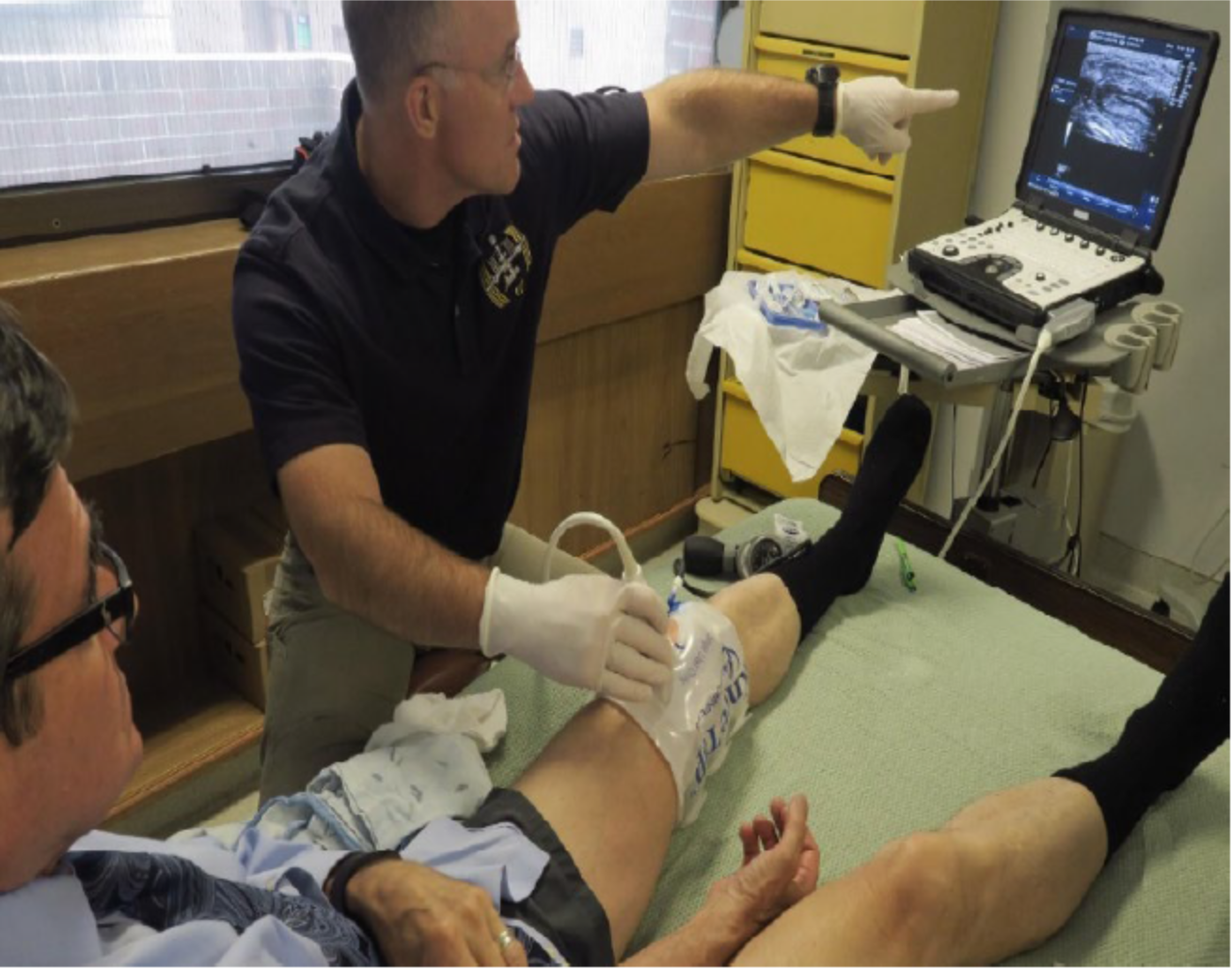
Image below, NASA/ JSC Flight Surgeon Dr Rick Scheuring performing knee ultrasound examination with KneeTapTM external pneumatic compressive device on RM at National Jewish Health Denver CO. (CREDIT: International Journal of Clinical Rheumatology)
Eighteen days in orbit did not appear to leave a measurable mark on the hips, knees, or ankles of three astronauts from Axiom Mission 4, according to new research from National Jewish Health. The result is reassuring on its own. Just as important, the work points to a portable imaging tool. This could help doctors keep closer watch on joint health during future missions. This is especially relevant as space agencies look toward longer stays in orbit and eventual trips to the Moon and Mars.
That tool is musculoskeletal ultrasound, a noninvasive scan more often associated with clinics on Earth than with astronaut medicine. In this pilot study, rheumatologists used it to examine cartilage, synovial fluid, tendons, and ligaments before launch. They also examined the astronauts again within four hours after splashdown. They found no statistically significant changes in the astronauts’ lower-extremity joint structures after the short mission aboard the International Space Station.
For a field concerned about what microgravity does to the human body, that matters.
Scientists have long known that reduced loading in space can weaken bone and shrink muscle. Joints raise a different concern. Cartilage depends on regular mechanical loading to stay healthy, yet it has little capacity to repair itself once damaged. Because it has no sensory nerve supply, trouble may not become obvious until substantial loss has already occurred.
Researchers have also worried about the supporting parts of joints, including tendons and ligaments. These structures help keep movement stable after astronauts return to Earth’s gravity.
A closer look at what changed, and what did not
The National Jewish Health team, led by Richard Meehan and Smarika Sapkota, studied three crewmembers from the Ax-4 mission. Ultrasound images were taken before flight in Houston in April 2025. The mission launched on June 25, 2025, and returned on July 15, 2025. All three astronauts were scanned in Long Beach, California, within four hours of splashdown.
The investigators measured cartilage depth in the knees, ankles, and hips, along with knee synovial fluid and the thickness of the infrapatellar ligament and Achilles tendon. They also used power Doppler imaging. This method can detect blood-flow signals associated with active inflammation.
They did not find evidence of that inflammation in the knees, ankles, hips, or in the insertion sites of the infrapatellar ligaments and Achilles tendons. Tendon and ligament thicknesses remained stable. Additionally, cartilage measurements in the femoral, talus, and hip regions also showed no significant differences before and after flight.
Even the knee fluid findings were mostly quiet. Two of the six knees measured showed an increase in synovial fluid after flight, by 4.4% and 21%. The other four showed decreases ranging from 5% to 25%. Taken together, those shifts were not statistically significant.
“This study provides encouraging early evidence that short-duration spaceflight, combined with exercise and medical countermeasures, may help preserve joint health,” Meehan said. “Equally important, it demonstrates that ultrasound can serve as a powerful, real-time tool to monitor joint health in space.”
That last point may be the more durable one.
Why joint damage in space remains a concern
The calm post-flight scans do not erase the larger problem. Earlier research has linked spaceflight and unloading to injuries and structural changes in the body. The study notes a high rate of knee injuries among Space Shuttle crew. In that group, 19 of 94 astronauts required surgical knee interventions.
It also cites six U.S. astronauts from long-duration International Space Station missions who were referred for consideration of total hip or knee arthroplasty. In another retrospective analysis of 242 astronauts, 9% sustained shoulder injuries. Those injuries were positively associated with missions lasting longer than six months.
Ground-based and animal studies have added to the concern. MRI scans have detected cartilage loss within weeks of human immobility and during head-down tilt bed rest, a common analog for spaceflight. Moreover, rodent research has also found cartilage thinning and other damage under unloading conditions and during orbital flight.
Flight duration impact
So why did this group come back looking largely unchanged?
The authors offer several possibilities. Eighteen days aboard the ISS may simply have been too short to produce measurable structural changes. All three crewmembers reportedly used non-steroidal anti-inflammatory drugs during flight, which may have helped blunt inflammatory effects. Each astronaut also performed cycling exercise, though the total amount varied, from 180 to 270 minutes over the mission.
Sapkota said the findings should not be stretched too far.
“Although we did not observe measurable changes after 18 days, longer missions could present very different risks to cartilage and joint structures,” she said. “Our findings highlight the importance of continued research and the potential of ultrasound to guide personalized countermeasures for astronaut health.”
A small study, but one with unusual timing
This was a proof-of-concept pilot study, and its limitations are not subtle. The sample size was only three people. The astronauts also differed in exercise exposure and in prior lower-extremity injury history. All had experienced earlier joint injuries or interventions, including ACL repair, ankle ligament reconstruction, knee arthroscopy, hyaluronic acid knee injections, and mesenchymal stem cell injections. That makes broad conclusions difficult.
Another possible confounder came after splashdown. The astronauts wore below-knee compression stockings for about three hours before the post-flight ultrasound exams, part of a routine effort to reduce orthostatic intolerance. The researchers note that this might have influenced synovial fluid measurements.
Still, the timing of the scans gave the work unusual value. Post-flight imaging happened quickly, before any temporary changes had much time to fade during recovery on Earth. The study also appears to be among the first to use quantitative ultrasound immediately after spaceflight. This was done to examine multiple lower-extremity joint structures in humans.
That speed matters because some biological changes can be fleeting. If clinicians want to catch them, they need tools that are fast, portable, repeatable, and practical.
Ultrasound fits that description better than MRI or CT. It does not use ionizing radiation, can be repeated in real time, and can be performed without the cost and logistical burden of larger imaging systems. In this study, it was paired with an external pneumatic compression device called KneeTapTM. This device is designed to help redistribute knee synovial fluid for more precise measurements.
Practical implications of the research
The most immediate implication is not that short missions are risk-free. It is that space medicine may have a workable way to monitor joint health without waiting for symptoms to appear.
For long-duration missions, that could be a big shift. Astronauts heading to the Moon or Mars will face much longer exposure to microgravity, and they will need ways to detect subtle joint changes before they become disabling. The researchers suggest future studies should include missions lasting three to six months. Ideally, these should use in-flight ultrasound exams and links to blood markers of inflammation.
They also point toward more advanced handheld ultrasound systems and even AI-guided probe placement to improve consistency from scan to scan.
The promise does not stop at the edge of space. Meehan said the same technology could help protect patients on Earth, including people recovering from prolonged immobility or those at risk of joint degeneration. Emmanuel Hilaire of National Jewish Health put it more broadly. He described space as a useful laboratory for developing biomedical tools with terrestrial value.
“This technology has the potential to transform how we monitor and protect joint health, not only for astronauts, but for patients here on Earth,” Meehan said.
Research findings are available online in the journal International Journal of Clinical Rheumatology.
The original story "Researchers find no significant joint damage in astronauts after short spaceflights" is published in The Brighter Side of News.
Related Stories
- Why do astronauts still act like gravity exists in space?
- Space travel accelerates stem cell aging - threatening astronaut health
- Flexible wearables revolutionize astronaut health monitoring in space
Like these kind of feel good stories? Get The Brighter Side of News' newsletter.
Mac Oliveau
Writer
Mac Oliveau is a Los Angeles–based science and technology journalist for The Brighter Side of News, an online publication focused on uplifting, transformative stories from around the globe. Having published articles on MSN, and Yahoo News, Mac covers a broad spectrum of topics including medical breakthroughs, health and green tech. With a talent for making complex science clear and compelling, they connect readers to the advancements shaping a brighter, more hopeful future.