Tiny spinning probe brings 360-degree view inside arteries
A 0.55-mm optical probe imaged stents and plaque while reducing rotational distortion inside curved cerebral vessels.

 Edited By: Joseph Shavit

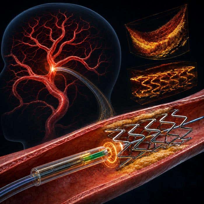
A 0.55-mm optical probe imaged stents and plaque while reducing rotational distortion inside curved cerebral vessels. (CREDIT: Dawei Wu, Nanjing University of Aeronautics and Astronautics)
- A 0.55-millimeter-wide optical probe traveled through a model of the brain’s blood vessels and produced full 360-degree images from inside them.
- A tiny piezoelectric actuator spins the lens directly at the catheter tip, reducing the rotational distortion that can occur when conventional catheters twist through curved arteries.
- The experimental device imaged stents, animal vessels and human plaque tissue, but it still needs improvements in resolution, stability, size and clinical safety testing.
A probe barely wider than half a millimeter has navigated a model of the human brain’s blood vessels while producing full 360-degree optical images. Its design could eventually give doctors a closer look inside small, twisting cerebral arteries.
The device measures 0.55 millimeters in diameter and 4 millimeters long. Researchers from Nanjing University and Nanjing University of Aeronautics and Astronautics developed it for intravascular optical coherence tomography, or OCT.
OCT uses light to create cross-sectional images of tissue. Inside blood vessels, it can reveal vessel walls, plaques and implanted devices such as stents.
The work showed that the probe could travel through a full-scale cerebrovascular model to the middle cerebral artery.
“Doctors currently have limited tools for looking at microscopic details directly inside small and tortuous brain arteries,” said Rui Liu of Nanjing University.
A motor at the catheter tip solves a twisting problem
Most intravascular OCT systems were designed for coronary arteries, which are generally larger and less tortuous than cerebral vessels.
Those systems usually rotate the imaging probe from outside the body. Rotation travels through a long catheter before reaching the tip.
In sharply curved vessels, friction and twisting can make that motion uneven. The resulting imaging error is called nonuniform rotational distortion, or NURD.
In tortuous vessels, the distortion can produce angular errors as large as 9 degrees.
Putting a motor directly at the catheter tip offers another option, but conventional electromagnetic micromotors become difficult to shrink below about 1 millimeter. Their wires can also block part of the optical field of view.
The new probe uses a piezoelectric actuator instead.
At its center is an optical fiber carrying near-infrared light to a tiny angled lens. Only that lens rotates, rather than the entire catheter.
A slanted groove in the probe converts back-and-forth vibration into twisting motion, spinning the lens around the vessel.
“We wanted to develop a fundamentally different miniature drive mechanism that could be placed at the distal tip of the catheter, remain extremely small, and still provide an unobstructed 360-degree optical scan,” Liu said.
The lens spins up to 58 times each second
Computer simulations helped the team choose the probe’s geometry and operating conditions. The final lens measured 0.185 millimeters in diameter and 1 millimeter long.
At 80 volts peak-to-peak, the lens reached 58 revolutions per second.
Researchers placed the device inside a 0.6-millimeter-diameter catheter and sent it through a full-scale vascular model.
Starting at the femoral artery, the catheter traveled through the abdominal aorta and carotid artery before reaching the middle cerebral artery.
The team then tested whether the rotational scan stayed uniform after that trip.
Four 1-millimeter metal tubes were arranged at known angles. After the catheter reached the middle cerebral artery, the probe imaged the tubes while rotating at 50 revolutions per second.
The measured angular deviation was only about 1 degree.
Optical tests reveal current resolution limits
The researchers measured imaging resolution with tungsten wires just 5 micrometers in diameter.
At a depth of 1 millimeter, lateral resolution reached 53.41 micrometers.
It declined with distance, measuring 80.18 micrometers at 1.8 millimeters and 119.68 micrometers at 3 millimeters.
The researchers concluded that the current probe works best for structures within about 2 millimeters in diameter.
They next tested increasingly complicated samples.
An air-dried magnolia leaf provided a network of vein-like structures. The probe distinguished a main vein about 1.8 millimeters across and secondary veins ranging from roughly 0.2 to 0.8 millimeters.
The device also scanned a stent expanded to a 2-millimeter inner diameter inside a middle cerebral artery model, producing a 3D reconstruction.
Vessels and human plaques add biological tests
The team also tested a porcine coronary vessel with an inner diameter of 2.5 millimeters. The images revealed areas of substantial lipid accumulation, although off-center positioning and light scattering weakened some signals.
Researchers then examined human atherosclerotic plaque removed during carotid surgery from a 60-year-old man.
OCT imaging showed collagen fibers spanning about 235 degrees of the plaque, with a maximum thickness near 109 micrometers.
The images also showed intimal thickening, tissue protruding into the vessel, a fibrous cap and a lipid-rich necrotic core.
A vascular pathologist who did not see the OCT results examined the tissue independently. Histology agreed with structures visible in the optical images, including plaque rupture sites and collagen-rich regions.
Clinical use will require more engineering
The device remains experimental.
Measured resolution was worse than simulations predicted, partly because of manufacturing errors, fiber-to-lens alignment and optical distortion from the protective sheath.
Fast rotation also caused minor lens wobbling, contributing to beam divergence and incomplete signals in some 3D images.
The researchers want to make the system smaller, improve resolution and imaging depth, and increase rotational stability and speed.
They believe the probe diameter could eventually shrink from 0.55 millimeters to around 0.4 millimeters.
Practical implications of the research
A smaller, tip-driven OCT probe could give neurointerventional physicians more detailed information about cerebral arteries than current intravascular systems can provide.
One possible use is checking how a stent sits against the vessel wall after placement.
“For example, it could provide a detailed view of how a stent is positioned relative to the vessel wall, potentially helping doctors make better treatment decisions and provide safer, more personalized care,” said Dawei Wu of Nanjing University of Aeronautics and Astronautics.
The technology could also help researchers study plaque structure and other microscopic features inside cerebral vessels.
Before clinical use, however, the probe must undergo further testing for imaging performance, reliability and safety under conditions that more closely resemble actual neurointerventional procedures.
For now, the experiments show that a piezoelectric motor can fit at the tip of an exceptionally small OCT catheter, keep its 360-degree scan uniform through tortuous paths and produce detailed images of stents and vascular tissue.
Dig deeper into optical imaging inside brain arteries
These resources explore the development of neurovascular OCT, its clinical potential and the challenge of imaging microscopic structures inside the brain’s tortuous arteries.
Volumetric microscopy of cerebral arteries with a miniaturized optical coherence tomography imaging probe: Reports a human study using a miniaturized neurovascular OCT probe to image cerebral arteries, vascular disease and implanted devices during endovascular procedures. (Science Translational Medicine, 2024)
A neurovascular high-frequency optical coherence tomography system enables in situ cerebrovascular volumetric microscopy: Describes an earlier neurovascular OCT system engineered to image arterial walls and implants within the difficult anatomy of cerebral blood vessels. (Nature Communications, 2020)
Development, image interpretation, clinical experience, and applications of optical coherence tomography in neurointerventional surgery: Reviews neuro-specific OCT development, early clinical experience and the principles needed to interpret high-resolution images of intracranial arteries and devices. (Journal of NeuroInterventional Surgery, 2026)
Optical coherence tomography in neurovascular intervention: Technical aspects and emerging directions: Examines the engineering requirements, limitations and potential applications of OCT for cerebral aneurysms, atherosclerosis, stroke and neurovascular implants. (Diagnostic and Interventional Imaging, 2026)
Optical Coherence Tomography in Cerebrovascular Disease: Open up New Horizons: Reviews the use of OCT for evaluating intracranial atherosclerosis, aneurysms, plaques and the results of stent treatment. (Translational Stroke Research, 2022)
Research findings are available online in the journal Biomedical Optics Express.
The original story "Tiny spinning probe brings 360-degree view inside arteries" is published in The Brighter Side of News.
Related Stories
- New optical trick pulls hidden quantum signals out of background noise
- MIT and Harvard break quantum limit with world’s most accurate optical clock
- Researchers develop a wearable device for safe, noninvasive, optical brain imaging
Like these kind of feel good stories? Get The Brighter Side of News' newsletter.
Rebecca Shavit
Writer
Based in Los Angeles, Rebecca Shavit is a dedicated science and technology journalist who writes for The Brighter Side of News, an online publication committed to highlighting positive and transformative stories from around the world. Having published articles on MSN, AOL News, and Yahoo News, Rebecca's reporting spans a wide range of topics, from cutting-edge medical breakthroughs to historical discoveries and innovations. With a keen ability to translate complex concepts into engaging and accessible stories, she makes science and innovation relatable to a broad audience.