
Scientists discover eye color played a significant role in vertebrate evolution
Tiny pigment granules in fossil fish eyes mirror patterns in living species, revealing ancient clues to vision and raising questions about adaptation to land.
 UCC Writer: Eoin Hahessy

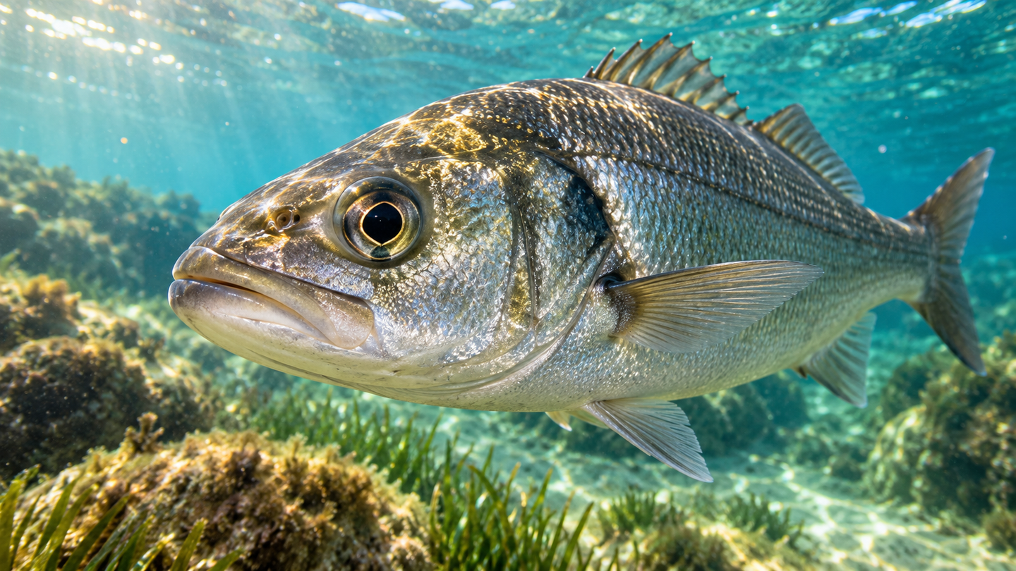
Microscopic pigment structures in fish eyes, also preserved in ancient fossils, offer clues to the evolution of vision. (CREDIT: Shutterstock)
- Microscopic pigment granules show distinct patterns across major fish groups, with matching features preserved in ancient fossils.
- Measurements of roughly 23,000 modern and 3,000 fossil melanosomes connect their shapes and locations with proposed visual adaptations.
- Lungfish pigmentation also suggests a role in coping with physiological stress, but its contribution to life on land remains a hypothesis.
Microscopic pigment granules inside fossil fish eyes preserve clues to how ancient animals handled light. Their shapes resemble those found in living fish, despite more than 300 million years separating the specimens. That continuity gives paleontologists a way to investigate aspects of sensory biology that bones alone cannot reveal.
A University College Cork-led study in Nature Communications links these structures, called melanosomes, with differences among major fish lineages. The researchers measured roughly 23,000 melanosomes from 12 living species and compared them with about 3,000 preserved in fossil fish. Their findings suggest that these granules record more than an animal's outward appearance. The collaboration included Shaun Collin at La Trobe University in Australia.
Melanosomes contain melanin, the pigment familiar from human skin and hair. Inside vertebrate bodies, melanin also has roles in regulating light and handling potentially harmful substances. The study examines how the size, shape and distribution of its storage structures relate to those functions, rather than treating pigment solely as a record of color.
Small structures with different jobs
The living species represented jawless fish, cartilaginous fish and the two major branches of bony fish. Examples included a river lamprey, sharks and skates, ray-finned fishes and a West African lungfish. Comparing their eyes, skin and internal tissues allowed the team to separate patterns associated with anatomy from those associated with evolutionary relationships.
Scanning electron microscopy revealed the tiny granules in detail, while statistical comparisons tested differences in their geometry. The researchers measured dimensions and compared how rounded or elongated the structures were. This approach matters because melanosomes from different tissues can overlap in shape, making simple visual descriptions less informative than measurements across many specimens.
Most internal tissues contained broadly rounded melanosomes with relatively limited variation. Eye granules showed much stronger differences among the groups examined. That contrast suggests that conditions inside the eye place distinctive demands on melanosome structure, including how pigment absorbs light and limits unwanted scattering.
Two ways to manage incoming light
The eyes of the sampled bony fish contained both rounded and elongated, cylindrical melanosomes. The researchers describe these informally as meatball-shaped and sausage-shaped structures. Jawless and living cartilaginous fish in the study had only the rounded population, providing a useful distinction among these broad evolutionary groups.
“These different melanosome shapes are linked to vision,” said lead author Dan Cirtina, a doctoral researcher at University College Cork. In bony fish, the elongated structures occur in a pigmented layer beside the retina's light-sensitive cells. Their arrangement and shape are consistent with reducing stray light and helping regulate exposure, potentially supporting sharper vision.
Many bony fish adjust to changing illumination partly by repositioning pigment and light-sensitive structures within the retina. Elongated melanosomes can move into narrow cellular projections, helping shield photoreceptors in bright conditions. These retinomotor movements can be slow, sometimes taking hours, so they offer a different solution from rapidly changing pupil size.
Sharks and rays commonly rely more heavily on pupil adjustments and adaptations that favor detecting limited light. In the examined cartilaginous fishes, pigment granules in the retinal pigment layer were reduced or absent, although melanosomes remained elsewhere in their eyes. That distinction is important: the study does not show that sharks lack eye pigment altogether.
Reducing pigment in that location can leave a reflective layer, called the tapetum lucidum, less obstructed. Reflected light gets another opportunity to reach photoreceptors, supporting sensitivity in dim conditions. The researchers interpret these arrangements as different balances among visual detail, light detection and adjustment speed, rather than a simple ranking of better and worse eyes.
Fossils preserve an ancient pattern
The fossil specimens came from the Mazon Creek biota in Illinois, a Carboniferous deposit approximately 306 million years old. Their preserved eye melanosomes broadly matched the patterns identified in living groups. Fossil jawless fish had rounded granules, while fossil bony fish also preserved elongated forms.
An extinct group called acanthodians likewise preserved cylindrical eye melanosomes. Their position near the ancestry of modern cartilaginous fishes supports the interpretation that elongated structures were present early in jawed vertebrate evolution. The team proposes that these structures were subsequently lost along the lineage leading to living sharks, rays and their relatives.
Distinguishing fossil evidence from evolutionary inference matters here. The specimens directly demonstrate the pattern at least 306 million years ago. Comparisons across the vertebrate family tree suggest that associated retinal adaptations could extend back toward the ancestors of jawed vertebrates around 445 million years ago, rather than documenting them directly in fossils that old.
Preservation also complicates the interpretation of size. Previous experiments show that melanosomes can shrink during heating associated with fossilization, meaning preserved dimensions may differ from those in living animals. The broad shape patterns remain informative, but reconstructing the original size and precise visual performance of an ancient eye requires additional evidence.
Lungfish hint at protection beyond pigment
The West African lungfish stood out for having larger melanosomes in several internal tissues and pigmentation across more internal organs. These tissues receive little direct light, making protection from sunlight an unlikely explanation for the pattern. Instead, the researchers propose a connection with the physiological demands of surviving seasonal drought.
African lungfish can enter aestivation, a dormant state that allows them to endure prolonged dry conditions with greatly reduced metabolism. Returning to higher activity can generate reactive oxygen species, chemically reactive molecules capable of damaging tissues. Melanin's known capacity to help neutralize such molecules makes extensive internal pigmentation a plausible part of the protective response.
However, this study did not experimentally demonstrate that those granules prevent stress damage or enabled ancient vertebrates to colonize land. The proposed link draws on comparative anatomy and existing knowledge of lungfish physiology. It raises a testable possibility about coping outside water, while limited fossil sampling and preservation leave room for alternative explanations.
Reading biology from ancient granules
“Our findings show that melanin is much more than a pigment used to colour our skin and hair,” said senior author Maria McNamara, professor of paleontology at University College Cork. The results connect microscopic anatomy with broader questions about vision and environmental adaptation. They also suggest that melanosome measurements could help classify fossils when more familiar anatomical features are missing.
That potential depends on expanding the comparison beyond the 12 living species examined here. Thousands of measured granules provide detailed information, but they do not represent thousands of independent species or encompass all fish diversity. A broader sampling could test how reliably these patterns distinguish lineages and how ecological differences modify them.
The findings provide a framework for investigating ancient biology without assuming every function can be read directly from a fossil. Shape, position and evolutionary context together offer stronger clues than any single granule. Further work can test whether the proposed visual and stress-protection roles explain the patterns across a wider range of living and extinct fish.
Dig deeper into melanosomes and vertebrate evolution
These resources connect fossil pigment evidence with visual adaptation, immunity and the evolution of melanin's functions.
Four camera-type eyes in the earliest vertebrates from the Cambrian Period
Fossil melanosomes help researchers interpret the visual organs of very early vertebrates, extending the context beyond Carboniferous fish. (Nature, January 2026)
Adaptations of the Vertebrate Retina to Low-Light Conditions: A Review
This review explains how reflective layers, photoreceptors and other retinal features balance light sensitivity against visual resolution. (Anatomia, Histologia, Embryologia, May 2025)
The melano-macrophage: The black leukocyte of fish immunity
The authors review pigment-containing immune cells and the roles of melanin beyond coloration, including responses to reactive molecules. (Fish & Shellfish Immunology, March 2024)
Research findings are available online in the journal Nature Communications.
The original story "Scientists discover eye color played a significant role in vertebrate evolution" is published in The Brighter Side of News.
Related Stories
- Mass extinction helped jawed vertebrates rise, study finds
- Scientists discover why animals evolved necks, and surprisingly it wasn’t to turn their heads
- Scotland’s famous 'Lizzie' fossil was not the early reptile scientists thought
Like these kind of feel good stories? Get The Brighter Side of News' newsletter.
Hannah Shavit-Weiner
Medical & Health Writer
Hannah Shavit-Weiner is a Los Angeles–based medical and health journalist for The Brighter Side of News, an online publication focused on uplifting, transformative stories from around the globe. Having published articles on AOL.com, MSN and Yahoo News, Hannah covers a broad spectrum of topics—from medical breakthroughs and health information to animal science. With a talent for making complex science clear and compelling, she connects readers to the advancements shaping a brighter, more hopeful future.