Adult brain cells reveal a surprising ability to repair damaged tissue
Adult astrocytes stretched, divided, and moved new nuclei into damaged brain regions during a temporary repair state.
 University of Zurich Writer: Kurt Bodenmueller

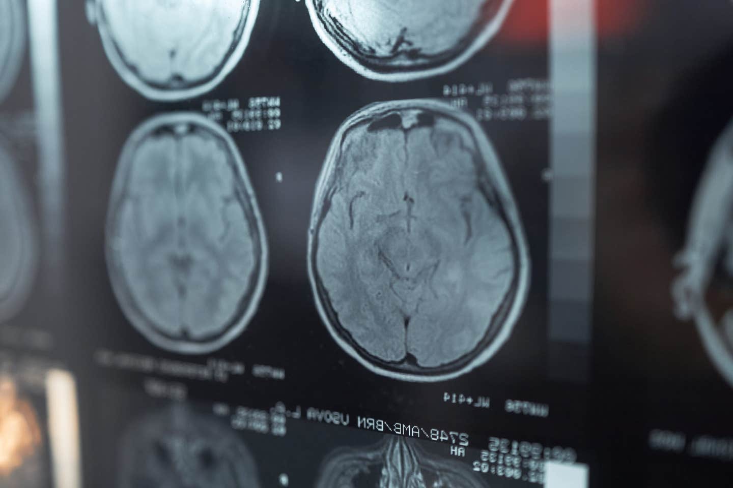
Adult astrocytes stretched, divided, and moved new nuclei into damaged brain regions during a temporary repair state. (CREDIT: Shutterstock)
A damaged patch of adult brain tissue can trigger nearby support cells to stretch, divide, and send newly formed nuclei into the empty space, revealing a repair response more flexible than scientists had assumed.
Researchers at the University of Zurich observed this process in living mice after selectively destroying groups of astrocytes, star-shaped glial cells that help support neurons and maintain healthy brain tissue.
The study showed that surviving astrocytes did not simply form a boundary around the damage. Many entered a temporary regenerative state, expanded their reach, divided, and gradually repopulated the depleted area.
The team also found astrocytes with some similar features in human brain tissue from people with neuromyelitis optica spectrum disorder, or NMOSD.
“The findings of our study reveal a previously unknown ability of the adult brain to repair itself,” said Bruno Weber, who led the research. “They point toward new ways of supporting recovery from ailments involving the loss of astrocytes.”
Astrocytes Move Into Emptied Brain Territory
Astrocytes perform several essential jobs in the brain. They provide nutrients to neurons, help regulate blood flow, and support the local environment around nerve cells.
Researchers had long assumed that adult brains could not fully replace astrocytes once large numbers were lost.
To test that idea, the team created small regions of astrocyte loss in the somatosensory cortex of adult mice. They used antibodies against aquaporin-4, or AQP4, a water channel found mainly on astrocytes in the central nervous system.
AQP4 antibodies also occur in NMOSD, an autoimmune disease in which astrocytes can come under attack.
Using long-term two-photon microscopy, the researchers repeatedly watched the same cells in living mice for as long as 60 days.
Astrocyte coverage gradually returned. Cell bodies reappeared inside depleted regions, and their numbers reached a plateau within several weeks.
The damaged areas also differed from some stroke or traumatic brain injury lesions because they did not develop a glial border that blocked astrocytes from entering.
Surviving Cells Stretch, Then Begin Dividing
Changes began quickly around the edge of the depleted areas.
Astrocyte extensions lengthened and pointed toward the center of the damaged region. Those processes reached their greatest length about five days after injury.
By the same point, the territory occupied by individual astrocytes had expanded by roughly 70 percent compared with before the lesion.
The cells were not merely stretching.
About 60 percent of astrocytes surrounding the lesion divided during the 60 days after injury. In control areas, only about 1 percent did so.
Some astrocytes underwent several rounds of division and produced as many as four daughter cells.
A separate labeling experiment supported that result. Roughly 50 to 60 percent of astrocytes around the injury showed signs of recent DNA replication by day 14.
Together, the observations indicated that mature cortical astrocytes retained a substantial ability to proliferate after nearby cells disappeared.
Daughter Nuclei Travel Through the Mother Cell
The most unusual part of the repair process came after division.
Instead of immediately separating into independent daughter cells, some astrocytes temporarily contained multiple nuclei.
Those newly formed nuclei then traveled through long extensions of the mother cell toward vacant regions.
“They send the newly formed nuclei of their daughter cells gliding across long distances to repopulate the damaged area of the brain and knit the astrocyte network back together,” Weber said.
Most movement occurred during the first two weeks.
About 75 percent of daughter nuclei relocated during the first seven days. More than 60 percent traveled farther than 30 micrometers from the mother cell.
Researchers recorded movements spanning more than 100 micrometers in some cases.
The nuclei did not travel smoothly. Imaging awake mice every three hours revealed a stop-and-start movement pattern through the astrocyte extensions.
Maximum movement speed reached 3.5 micrometers per hour, with peak rates of 5.6 micrometers per hour. Some nuclei remained still for as long as nine hours.
As they moved, the nuclei stretched into elongated shapes and later became rounder again.
A Temporary Genetic Repair Program Switches On
The physical changes coincided with a major shift in gene activity.
Researchers mapped which genes became more or less active at several points after injury. Thousands changed their activity during the early stages of repair.
Genes associated with cellular plasticity and structural remodeling became especially active.
That repair state peaked around day five, then faded. By day 17, the injury-associated genetic profile was no longer clearly detectable as a separate state.
Calcium signaling showed a similarly temporary response.
By day five, astrocytes near the lesion displayed stronger and more frequent calcium activity. By day 14, those patterns had returned toward normal as the damaged region filled back in.
“We were able to identify numerous genes and signaling pathways that are temporarily activated during repair,” Weber said. “They could serve as starting points in the future for influencing post-disease and -injury regeneration processes.”
The Type and Size of Damage Changed the Response
Astrocytes did not regenerate equally well after every kind of injury.
When the researchers created small stroke-like lesions, astrocytes still divided, but repair remained incomplete. Nuclear movement was less common and covered shorter distances.
The size of astrocyte loss also mattered.
Destroying only a few astrocytes triggered little response. Removing a thin layer caused neighboring cells to stretch toward the damage but produced little proliferation.
Larger three-dimensional losses triggered the strongest response, including polarization, division, and long-distance nuclear movement.
Those results indicate that adult astrocyte repair depends on the nature and extent of the damage.
Human Tissue Offers a Cautious Parallel
The team also examined seven human brain samples from confirmed AQP4-antibody-positive NMOSD cases.
Affected areas contained astrocytes that were elongated, oriented toward lesions, sometimes multinucleated, and in some cases showed markers of proliferation.
Those cells appeared more often in affected tissue than in unaffected areas.
However, the researchers cautioned that human NMOSD involves chronic inflammation, immune activity, and broader tissue damage that the mouse model does not reproduce.
The human samples therefore cannot establish that patients undergo the same repair process seen in mice.
Practical Implications of the Research
The findings provide a new framework for studying how adult astrocytes respond when neighboring cells disappear.
Future work could test whether the temporary repair program can be safely strengthened or prolonged in conditions involving astrocyte loss.
The genes and signaling pathways activated during regeneration offer possible targets for that research, but the current experiments do not establish a treatment.
The work also suggests that brain repair cannot be described by a single rule. Astrocytes reacted differently depending on injury type, lesion size, and whether a restrictive border formed.
Understanding those conditions may help researchers determine when the adult brain can rebuild astrocyte networks on its own and when that process fails.
Research findings are available online in the journal Nature Neuroscience.
The original story "Adult brain cells reveal a surprising ability to repair damaged tissue" is published in The Brighter Side of News.
Related Stories
- Stanford researchers discover a language-specific network hidden in the human brain
- Generations of breeding changed how dogs’ brains process human communication
- MIT study finds hidden language network in the human brain
Like these kind of feel good stories? Get The Brighter Side of News' newsletter.
Joshua Shavit
Writer and Editor
Joshua Shavit is a NorCal-based science and technology writer with a passion for exploring the breakthroughs shaping the future. As a co-founder of The Brighter Side of News, he focuses on positive and transformative advancements in technology, physics, engineering, robotics, and astronomy. Having published articles on AOL.com, MSN, Yahoo News, and Ground News, Joshua's work highlights the innovators behind the ideas, bringing readers closer to the people driving progress.